VISE Seminar: High Throughput Histological and Metabolic Imaging of Surgical Specimens
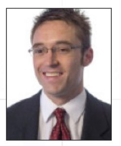
Speaker: Michael G. Giacomelli
Postdoctoral Fellow,
Biomedical Optical Imaging and Biophotonics Group
Department of Electrical Engineering and Computer Science
Research Lab of Electronics, Massachusetts Institute of Technology
Date: Thursday, January 19, 2017
Time: 12:20 start, lunch 12:15
Place: Stevenson Center 5326
Title: High throughput histological and metabolic imaging of surgical specimens
Abstract: Optical imaging of preserved, thinly sectioned tissue is the standard for the diagnosis of most cancers. However, in spite of revolutionary advances in scientific imaging and computer science, many widely used histological techniques have changed little since the late 19th century. Virtual histology, which combines computational methods with imaging techniques such as nonlinear, confocal, fluorescent lifetime, and UV microscopy to produce real-time digital histology images of tissue, has the potential to improve surgical treatment, reduce medical costs, and provide wider access to care. Novel developments in high throughput histological imaging, miniaturization, processing and visualization will be presented in the context surgical imaging and cancer diagnosis.
Bio: Michael Giacomelli received dual bachelor’s degrees from the University of Arizona in Computer Science and Computer Engineering in 2006, and a Ph.D. in Biomedical Engineering from Duke University in 2012. He is currently an NIH NRSA postdoctoral fellow in the Research Laboratory of Electronics at MIT. His research interests include surgical and endoscopic imaging using nonlinear, confocal, and fluorescent lifetime microscopy, as well as interferometry, computational approaches to light scattering and inverse problems, and digital pathology.

