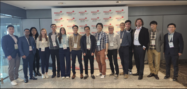
VISE on the Road: 2023 MICCAI conference

Members of six VISE labs affiliated with the Vanderbilt Institute for Surgery and Engineering (VISE) traveled to Vancouver to take part in the 26th International Conference on Medical Image Computing and Computer-Assisted Intervention. Like-minded scientists from around the world gather yearly at the MICCAI conference to share their work with one another on a grand stage.
The annual conference attracts leading biomedical scientists, engineers, and clinicians from multiple disciplines associated with medical imaging and computer-assisted intervention. The conference includes oral presentations, poster sessions, workshops, tutorials, and challenges. The VISE labs and presenters were:
Medical-image Analysis and Statistical Interpretation Lab (MASI)
Bennett Landman, PhD, Chair, Department of Electrical and Computer Engineering, co-lead the QuantConn challenge with CDMRI, the International Workshop on Computational Diffusion MRI.
“MICCAI is a fantastic venue to share and discuss deep technical innovations,” Landman said.
“In addition to the large center stage, we have developed a robust satellite community that brings together smaller groups to advance specialized science. This year, we led the QuantConn challenge as a satellite event to move toward using diffusion MRI tractography as biomarkers.”
Graduate student Nancy Newlin, one of the challenge organizers, gave a QuantConn talk titled “Introducing QuantConn: Overcoming challenging diffusion acquisitions with harmonization.”
“It was an honor to work as point person for this project and engage with these researchers firsthand,” she said. “I’m excited to publish the findings later this year!”
Thomas Li, a MD/PhD student, presented an oral talk titled, “Longitudinal Multimodal Transformer Integrating Imaging and Latent Clinical Signatures from Routine EHRs for Pulmonary Nodule Classification.”
During a poster session, graduate student, Peter Lee, shared his work titled “Scaling up 3D Kernels with Bayesian Frequency Re-parameterization for Medical Image Segmentation”
Biomedical Data Representational and Learning Lab (HRLB)
Assistant Professor of Electrical and Computer Engineering Yuankai Huo was happy to return to the first in-person MICCAI conference in North America since COVID.
“You could really feel the excitement in the air – everyone was so thrilled to be back, sharing ideas face-to-face,” he said.
“And the advancements, like in multi-modal and self-supervised learning in medical image analysis, were just the cherry on top. It felt like we were not just catching up on lost time but actually leaping forward. To be part of this, after such a challenging time, was really something special.”
Huo shared graduate student Tianyuan Yao’s work on diffusion harmonization with an oral presentation titled “A unified single-stage learning model for estimating fiber orientation distribution functions on heterogeneous multi-shell diffusion-weighted MRI.” Yao attended virtually.
Graduate student Ruining Deng won second place for his entry—”Knowledge-Infused Efficient Learning for Giga-Pixel Virtual Microscopy Images”—in the MICCAI Student Board Thesis Madness, a 3-minute PhD thesis competition.
Deng also presented his first author paper during the MICCAI main conference. Title: “Democratizing Pathological Image Segmentation with Lay Annotators via Molecular-empowered Learning.”
Deng enjoyed the in-person opportunity to ask questions and receive advice with experts in the field. “The biggest takeaway is understanding how to define a good question, one that can resolve clinical problems and have clinical value while also driving technical innovation. With such solutions, we can build a knowledge bridge between the medical/surgical/clinical fields and engineering,” he said.
Can Cui presented her first author paper at MILLAND workshop titled “Feasibility of Universal Anomaly Detection Without Knowing the Abnormality in Medical Images.”
She said the conference was a fantastic experience and cited connecting with old and new friends as a highlight. “I had the opportunity to engage in face-to-face discussions with other researchers and witness the enthusiasm among researchers for various exciting topics in the medical image domain,” she said.
Quan Liu presented his first author paper (remotely) during the MMMI workshop: “M^2Fusion: Bayesian-based Multimodal Multi-level Fusion on Colorectal Cancer Microsatellite Instability Prediction.”
“I had the opportunity to immerse myself in a series of captivating talks, each centered around distinct topics such as multimodal learning, efficient learning, and histopathology image analysis,” Liu said. “The ideas presented were truly inspiring, and the cutting-edge methods discussed have left me feeling not only motivated but also well-equipped for my future research endeavors.”
Medical Image Computing Lab (MedICL)
Two students in the MedICL lab, under the direction of Assistant Professor of Computer Science Ipek Oguz, won awards during the conference.
Han Liu’s team won the CrossMoDA challenge with their project titled: “Learning Site-specific Styles for Multi-institutional Unsupervised Cross-modality Domain Adaptation.” In addition, Liu presented ““COLosSAL: A Benchmark for Cold-start Active Learning for 3D Medical Image Segmentation” in a workshop.
David Lu won the outstanding paper award at the AE-CAI workshop. His paper is titled: “ASSIST-U: A System for Segmentation and Image Style Transfer for Ureteroscopy.” This was Lu’s first, first-author paper and he said, “It felt incredibly encouraging to be recognized.” Lu also presented a workshop talk on titled “MAP: Domain Generalization via Meta-Learning on Anatomy-Consistent Pseudo-Modalities” on behalf of Dewei Hu at MedAGI.
Visual Informatics and Engineering Lab (VINE)
Assistant Professor of Computer Science Daniel Moyer organized two satellite events, the Fairness in AI and Medical Imaging (FAIMI) workshop, and a tractography challenge with Bennett Landman’s students/collaborators.
Machine Automation, Perception, and Learning Lab (MAPLE)
Graduate students John Han, Ayberk Acar, and Jumanh Atoum, along with research assistant Yinhong Quin, won Best Methodology Report award in the Surgical Tool Localization in Endoscopic Videos, which was part of the EndoVis challenge. The MAPLE lab is under the direction of assistant professor in computer science Jie Ying Wu.
Graduate student Ayberk Acar presented his first author paper during the AE-CAI | CARE | OR 2.0 workshop titled: “Towards Navigation in Endoscopic Kidney Surgery based on Preoperative Imaging.” He also presented additional papers in that workshop for students who were unable to attend.
First authors Ayberk Acar and Jumanh Atoum paper title: “Intraoperative Gaze Guidance with Mixed Reality”
First authors Guansen Tong and Jiayi Li paper title: “Development of an Augmented Reality Guidance System for Head and Neck Cancer Resection”
Biomedical Image Analysis for Image Guided Interventions Lab (BAGL)
Graduate student Mohammad Khan presented his poster and fellow lab mate Ziteng Liu’s paper during the Simulation and Synthesis in Medical Imaging workshop. Khan’s poster title: “Cochlear Implant Fold Detection in Intra-operative CT using Weakly Supervised Multi-Task Deep Learning.” Liu’s paper: “Super-resolution segmentation network for inner-ear tissue segmentation.”
Khan found MICCAI an enriching experience. “The opportunity to engage with experts and learn about the latest developments in my field has equipped me with new algorithms and cutting-edge techniques. The experience has expanded my knowledge and will undoubtedly shape the future of my work,” he said.
Medical Image Processing Lab (MIP)
Graduate student Yubo Fan echoed Khan sentiment. “Attending MICCAI in person and presenting our work there was a fantastic experience,” he said.
“I was especially impressed by the exceptional quality of cutting-edge research showcased at the conference. It was really nice to see that there are many interesting problems to solve in this field. The opportunity to network with other researchers in academia and industry was also invaluable.”
Fan presented during a workshop and poster session. Workshop paper title: “A Unified Deep-Learning-Based Framework for Cochlear Implant Electrode Array Localization. ”Poster title “CT Synthesis with Modality-, Anatomy-, and Site-Specific Inference.”
The Vanderbilt Institute for Surgery and Engineering (VISE) is an interdisciplinary, trans-institutional structure designed to facilitate interactions and exchanges between engineers and physicians. Its goal is to become the premier institute for the training of the next generation of surgeons, engineers, and computer scientists capable of working symbiotically on new solutions to complex interventional problems, ultimately resulting in improved patient care.

