Labs
Labs
VISE Affiliated Labs
The Vanderbilt Institute for Surgery and Engineering (VISE) is an interdisciplinary, trans-institutional entity designed to facilitate these interactions and exchanges. Its mission is the creation, development, implementation, clinical evaluation and commercialization of methods, devices, algorithms, and systems designed to facilitate interventional processes and their outcome. Its goal is to become the premier center for the training of the next generation of surgeons, engineers, and computer scientists capable of working synergistically on new solutions to complex interventional problems, ultimately resulting in improved patient care. VISE includes ten technical laboratories spanning three engineering departments (Biomedical Engineering, Mechanical Engineering, and Electrical Engineering and Computer Science) and the Otolaryngology department as well as clinical departments that include Surgery, Neurological Surgery, Radiology, Otolaryngology, Hearing and Speech, Oncology, Gastroenterology, Surgical and Radiological Oncology, Ophthalmology, Urology, and Thoracic Surgery.
Advanced Robotics and Mechanism Applications (ARMA) Laboratory
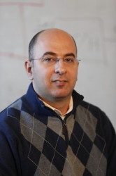
PI: Nabil Simaan, Professor of Mechanical Engineering & Otolaryngology
ARMA is focused on advanced robotics research including robotics, mechanism design, control, and telemanipulation for medical applications. We focus on enabling technologies that necessitate novel design solutions that require contributions in design modeling and control. ARMA has lead the way in advancing several robotics technologies for medical applications including high dexterity snake-like robots for surgery, steerable electrode arrays for cochlear implant surgery, robotics for single port access surgery and natural orifice surgery. Current and past funded research includes transurethral bladder cancer resection (NIH), trans-oral minimally invasive surgery of the upper airways (NIH), single port access surgery (NIH), technologies for robot surgical situational awareness (National Robotics Initiative), Micro-vascular surgery and micro surgery of the retina (VU Discovery Grant), Robotics for cochlear implant surgery (Cochlear Corporation). We collaborate closely with industry on translation our research. Examples include technologies for snake robots licensed to Intuitive Surgical, technologies for micro-surgery of the retina which lead to the formation of AURIS surgical robotics Inc., the IREP single port surgery robot which has been licensed to Titan Medical Inc. and serves as the research prototype behind the Titan Medial Inc. SPORT (Single Port Orifice Robotic Technology). Contact Nabil Simaan
View ARMA lab video here: ![]()
Biomedical Elasticity and Acoustic Measurement (BEAM) Laboratory

PI: Brett Byram, Hoy Family Faculty Fellow, Associate Professor of Biomedical Engineering
The biomedical elasticity and acoustic measurement (BEAM) lab is interested in pursuing ultrasonic solutions to clinical problems. Brett Byram and the BEAM lab’s members have experience with most aspects of systems level ultrasound research, but our current efforts focus on advanced pulse sequencing and algorithm development for motion estimation and beamforming. The goal of our beamforming work is to make normal ultrasound images as clear as intraoperative ultrasound, the gold-standard for many applications. We have recently demonstrated non-contrast tissue perfusion imaging with ultrasound at clinical frequencies, and we are developing novel ultrasound transducers to enhance guidance for percutaneous procedures.
Contact Brett Byram
Biomedical Image Analysis for Image Guided Interventions (BAGL) Laboratory
PI: Jack H. Noble, Assistant Professor of Electrical and Computer Engineering
Biomedical image analysis techniques are transforming the way many clinical interventions are performed and enabling the creation of new computer-assisted interventions and surgical procedures. The Biomedical Image Analysis for Image-Guided Interventions Lab (BAGL) investigates novel medical image processing and analysis techniques with emphasis on creating image analysis-based solutions to clinical problems. The lab explores state-of-the-art image analysis techniques, such as machine learning, statistical shape models, graph search methods, level set techniques, image registration techniques, and image-based bio-models. The lab is currently developing novel systems for cochlear implant procedures including systems that use image analysis techniques for (1) comprehensive pre-operative surgery planning and intra-operative guidance and (2) post-operative informatics to optimize hearing outcomes. Contact Jack Noble
Biomedical Modeling Laboratory (BML)
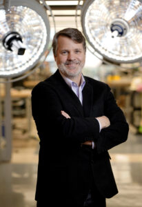
PI: Michael I. Miga, Chair of Biomedical Engineering, Harvie Branscomb Professor, Professor of Biomedical Engineering, Director of VISE
While research in diagnostic imaging, therapeutics, and drug delivery is quite extensive, advances in procedural medicine have lagged, largely due to the complexity and constraints associated with surgical and interventional research. However, with the recent breakthroughs in medical robotics, computer vision, artificial intelligence, and device fabrication, new opportunities are on the horizon for creating platform technologies characterized by an intentional design toward the dual purpose of treatment and discovery. Projects in the Biomedical Modeling Laboratory (BML, https://migalab.org/) center on complex biophysical models, methods of automation and procedural field surveillance, efforts toward data-driven procedures and therapy forecasting, and approaches integrating disease information associated with digital twins. While it is difficult to be prescient on the forms that these forward-thinking platforms will take in precision medicine, it is clear that new requirements associated with data integration/acquisition, automation, real-time computation, novel device development, and cost will likely be critical factors. Technologies will need to be enabling, efficient, and cost-effective and for medical systems that do not adapt precision care models, they will be left behind. Similarly, immersive training environments will be critical for working at the cutting-edge. To achieve, BML PI Dr. Mike Miga has pioneered one of those most innovative immersive educational environments in the world for engineering researchers working in the fields of surgery and intervention. Contact Michael Miga
View BML lab video here: ![]()
Bowden Biomedical Optics (BBOL) Laboratory
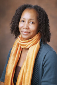 PI: Audrey Bowden, Dorothy J Wingfield Phillips Chancellor Faculty Fellow, Associate Professor, Biomedical Engineering, Associate Professor, Electrical and Computer Engineering
The primary aim of the Bowden Biomedical Optics Laboratory (BBOL) is to develop and deploy novel imaging and sensing technologies to address unmet clinical needs in medicine and biology.
We blend knowledge and experience from diverse fields such as optics, microfluidics, signal processing and computer science to develop software- and hardware-based tools for the healthcare provider that advance the state of the art and aid in scientific discovery. While the majority of our solutions are relevant to optics, as engineers, we are committed to taking a “whatever means necessary” approach to solving the clinical problem. We are also committed to developing novel solutions to improve delivery and affordability of healthcare in low-resource and resource-constrained environments. Our technologies and projects have found application in various clinical departments, including urology, dermatology, otolaryngology and women’s health. Contact Audrey Bowen
Brain Imaging and Electrophysiology Network (BIEN) Laboratory

PI: Dario Englot, Associate Professor of Neurological Surgery, Radiology and Radiological Sciences, and Biomedical Engineering
The BIEN lab integrates human neuroimaging and electrophysiology techniques to study brain networks in both neurological diseases and normal brain states. The lab is led by Dario Englot, a functional neurosurgeon at Vanderbilt. One major focus of the lab is to understand the complex network perturbations in patients with epilepsy, by relating network changes to neurocognitive problems, disease parameters, and changes in vigilance in this disabling disease. Multimodal data from human intracranial EEG, functional MRI, diffusion tensor imaging, and other tools are utilized to evaluate resting-state, seizure-related, and task-based paradigms. Other interests of the lab include the effects of brain surgery and neurostimulation on brain networks in epilepsy patients, and whether functional and structural connectivity patterns may change in patients after neurosurgical intervention. Through studying disease-based models, the group also hopes to achieve a better understanding of normal human brain network physiology related to consciousness, cognition, and arousal. Finally, surgical outcomes in functional neurosurgery, including deep brain stimulation, procedures for pain disorders, and epilepsy, are also being investigated. Contact Dario Englot
Diagnostic Imaging and Image-Guided Interventions (DIIGI) Laboratory
 PI: Yuankai (Kenny) Tao, Assistant Professor of Biomedical Engineering
The Diagnostic Imaging and Image-Guided Interventions (DIIGI) Laboratory develops novel optical imaging systems for clinical diagnostics and therapeutic monitoring in ophthalmology and oncology. Biomedical optics enable non-invasive subcellular visualization of tissue morphology, biological dynamics, and disease pathogenesis. Our ongoing research primarily focuses on clinical translation of therapeutic tools for image-guided intraoperative feedback using modalities including optical coherence tomography (OCT), which provides high-resolution volumetric imaging of weakly scattering tissue; and nonlinear microscopy, which has improved molecular-specificity, imaging depth, and contrast over conventional white-light and fluorescence microscopy. Additionally, we have developed optical imaging techniques that exploit intrinsic functional contrast for in vivo monitoring of blood flow and oxygenation as surrogate biomarkers of cellular metabolism and early indicators of disease. The majority of our research projects are multidisciplinary collaborations between investigators in engineering, basic sciences, and medicine. Contact Yuankai (Kenny) Tao View DIIGI lab video here: ![]()
Du Group
 PI: Yayun Du, Assistant Professor of Electrical and Computer Engineering
The lab focuses on advancing bioelectronics and intelligent systems through three key areas:
Wearable and Implantable Bioelectronic Sensors: We design, fabricate, and develop signal processing for multimodal, multi-point bioelectronic systems, with applications in intelligent health monitoring, disease diagnosis, and prevention, particularly in brain and cardiovascular science.
Human-in-the-Loop Interaction: We create safe and efficient systems for human-robot/computer interaction, leveraging bioelectronics and brain-computer interfaces to enhance functionality and user experience.
Adaptive Robotics and Automation: We integrate sensing, control, and machine learning (including reinforcement learning) to develop adaptive robotics for challenging medical environments, such as hospital wards and ICUs.
tHe biomedical data Representation and Learning laB
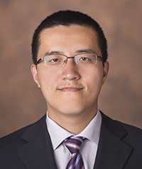 PI: Yuankai Huo, Assistant Professor in Computer Science
The HRLB lab aims to facilitate data-driven healthcare and improve patient outcomes through innovations in medical image analysis as well as multi-modal data representation and learning. Our current focus efforts on quantifying high-resolution and spatial-temporal data from microscopy imaging techniques, including renal pathology, cancer pathology, cytology, computational biology. The quantitative imaging information is associated with molecular, genetic, and clinical features for precise diagnosis and treatment. Contact Yuankai Huo
Internet of Medical Things (IoMT) Lab
 PI: James Weimer, Assistant Professor of Computer Science, Vanderbilt University
The internet of medical things (IoMT) consists of devices, infrastructure, and software connected through communication networks (e.g., the internet or hospital intranet). Consequently, in the past decade, the IoMT has grown to incorporate most commercial medical devices and consumer health products. In the IoMT lab, we seek to push the boundaries of how the IoMT can impact clinical care and patient health. The IoMT lab connects clinicians and engineers with the IoMT to create new inter-operable learning-enabled medical systems. Through collaborative interdisciplinary use-inspired research, we seek to address three foundational challenges facing the IoMT. First, the IoMT requires systems and protocols for identifying and collecting the right data in a timely manner. Second, the IoMT data must be processed to provide actionable feedback to clinicians and caregivers. Third, the IoMT should safely automate some aspects of care to reduce clinician and caregiver workload. To maximize the real-world IoMT lab impact, we go beyond traditional academic research and innovate — often developing intellectual property that is licensed to commercial entities. Through licensing and research, the IoMT lab has partnered with startup companies such as Neuralert and Vasowatch, as well as larger companies including Hill-Rom. Graduate and undergraduate students in the IoMT Lab have a unique educational experience that includes working side-by-side with clinicians. In the IoMT lab, students are encouraged to not only work on lab projects, but to pursue their own ideas as they learn to be both researchers and innovators in medical devices and health technologies. Contact James Weimer
Laboratory for the Design and Control of Energetic Systems (DCES)
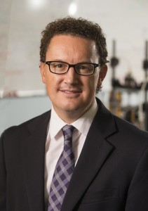 PI: Eric Barth, Professor of Mechanical Engineering, Professor of Neurological Surgery
Our Lab seeks to develop and experimentally apply a system-dynamics and control perspective to problems involving the control and transduction of energy. This is typically applied to problems in the fluid power domain (pneumatics and hydraulics) mixed with other energy domains (mechanical, thermal, etc). Our specific expertise in precision pneumatic control and mechanical design has enabled the realization of MRI compatible, pneumatically actuated, robotic platforms capable of accurate manipulation and force control for surgical tasks. Our lab is one of a handful in the world that have experimentally achieved control of pneumatic systems at the submillimetric accuracies needed for MRI compatible surgical systems. Pneumatic systems are highly nonlinear, high order dynamic systems and require sophisticated model-based nonlinear control for stability robustness, desirable dynamic response, and high accuracy positioning. Our lab also has an interest in designing and controlling pneumatic and hydraulic-powered soft robots for clinical and non-clinical applications. Most recent research efforts have focused on MRI compatible pneumatically actuated robotics, mechanical circulatory support devices including artificial hearts, and soft robotics, a new class of robot. Contact Eric Barth
Laboratory for Organ Recovery, Regeneration and Replacement (LOR3)
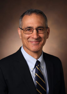
PI: Matthew Bacchetta,H. William Scott, Jr. Chair in Surgery, Associate Professor of Surgery
The LOR3 is focused on creating organ support systems that provide extended physiologic support for injured organs, bioengineering platforms for organ recovery and regeneration as well as developing artificial pulmonary assist devices. The lab maintains a full complement of devices used for extracorporeal life support and has developed durable support systems for lung and liver with translational potential. It works in partnerships with programs at VUMC, Carnegie Mellon University and Columbia University. The LOR3 is dedicated to translating basic science research into clinical platforms for patients with end organ failure.
Contact Matthew Bacchetta
Machine Automation, Perception, and Learning (MAPLE) Laboratory
 PI: Jie Ying Wu, Assistant Professor, Computer Science
At the MAPLE lab, our goal is to build intelligent surgical robots that can assist surgeons in the operation room. While surgical robots have changed many procedures by providing higher dexterity, motion scaling, and other innovations, they are still only extensions of the surgeon’s arms. By modeling different aspects of surgery and how they interact, we aim to make the robots more capable. We use machine learning to augment traditional modeling techniques, such as correcting physics-based soft-tissue models with observations of tissue interactions. We work with clinicians to use accurate soft-tissue models to provide guidance during surgeries based on preoperative imaging. Another project looks at modeling how expert surgeons move surgical instruments and the endoscope during procedures, which can help us develop better ways to train novice surgeons. At the same time, we are building models of the trainee’s actions, eye-gaze, and pupillometry to obtain insight into their cognitive load. We use this to develop a personalized curriculum and feedback. Contact Jie Ying Wu
Machine Intelligence and Neural Technologies (MINT) Laboratory
 PI: Soheil Kolouri, Assistant Professor of Computer Science
At the Machine Intelligence and Neural Technologies (MINT) Lab, we develop next-generation core Machine Learning (ML) solutions for practical problems in medicine and strive to advance healthcare. Our interdisciplinary team at MINT Lab uses biological inspirations together with mathematical and geometrical tools to innovate theoretically grounded algorithms that address the current deficiencies in ML technologies regarding lifelong/continual learning, sample/label efficiency, explainability, and brittleness. In one of our main research thrusts, we develop brain-inspired, robust machine intelligence that can continually learn and adapt to the input stream of nonstationary multimodal data. Continual learning is specifically relevant to medical applications where: 1) the data is continually accumulated from new patients, and 2) diseases constantly mutate and new variants emerge. We are developing next-generation computational models that adapt to these constant variations, learn from the past to solve future problems and leverage new knowledge to improve the previous solutions. Our research is highly interdisciplinary, and we have collaborations across fields including computer science, biomedical engineering, cognitive science, electrical engineering, and neuroscience. Contact Soheil Kolouri
Medical Engineering and Discovery (MED) Laboratory
 PI: Robert J. Webster III, Richard A. Schroeder Professor of Mechanical Engineering, Associate Provost for Graduate Education,
Professor of Mechanical Engineering
The Vanderbilt School of Engineering’s Medical Engineering and Discovery (MED) Laboratory pursues research at the interface of surgery and engineering. Our mission is to enhance the lives of patients by engineering better devices and tools to assist physicians. Much of our current research involves designing and constructing the next generation of surgical robotic systems that are less invasive, more intelligent, and more accurate. These devices typically work collaboratively with surgeons, assisting them with image guidance and dexterity in small spaces. Creating these devices involves research in design, modeling, control, and human interfaces for novel robots. Specific current projects include needle-sized tentacle-like robots, advanced manual laparoscopic instruments with wrists and elbows, image guidance for high-accuracy inner ear surgery and abdominal soft tissue procedures, and swallowable pill-sized robots for interventions in the gastrointestinal tract. Contact Robert Webster .
View MED lab video here: ![]()
Medical-image Analysis and Statistical Interpretation (MASI) Laboratory
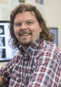
PI: Bennett Landman, Director, Vanderbilt Lab for Immersive AI Translation (VALIANT), Stevenson Chair, Professor of Electrical and Computer Engineering
Three-dimensional medical images are changing the way we understand our minds, describe our bodies, and care for ourselves. In the MASI lab, we believe that only a small fraction of this potential has been tapped. We are applying medical image processing to capture the richness of human variation at the population level to learn about complex factors impacting individuals. Our focus is on innovations in robust content analysis, modern statistical methods, and imaging informatics. We partner broadly with clinical and basic science researchers to recognize and resolve technical, practical, and theoretical challenges to translating medical image computing techniques for the benefit of patient care. Contact Bennett Landman
View MASI lab video here: ![]()
Medical Image Computing Laboratory (MedICL)
 PI: Ipek Oguz, Associate Professor of Computer Science
The goal of the Medical Image Computing Lab is to develop novel algorithms for better leveraging the wealth of data available in medical imagery. We are interested in a wide variety of methods including image segmentation, image registration, image prediction/synthesis, and machine learning. One of our current clinical applications is Huntington’s disease, where we are interested in improving the prediction of clinical disease onset through longitudinal segmentation of subcortical and cortical anatomy from brain MRI’s. We are also interested in multiple sclerosis, where we work on improving our understanding of both the inflammatory disease process through lesion quantification and a potential complementary neurodegenerative component through cortical thickness studies. Additional application areas include retinal OCTs and diffusion MRI in Aicardi-Goutières syndrome.
Contact: Ipek Oguz
Medical Image Processing (MIP) Laboratory
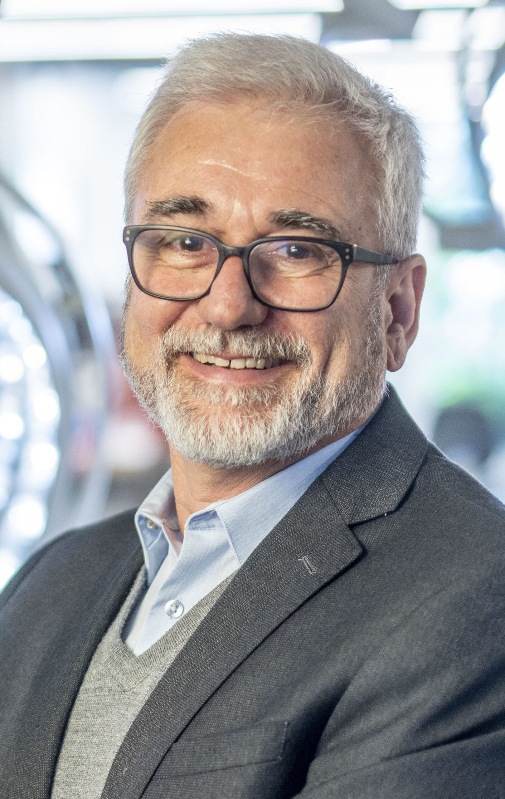 PI: Benoit Dawant, Interim Chair, Department of Electrical and Computer Engineering, Cornelius Vanderbilt Professor of Engineering, Professor of Electrical and Computer Engineering
The medical image processing (MIP) laboratory of the Electrical Engineering and Computer Science (EECS) Department conducts research in the area of medical image processing and analysis. The core algorithmic expertise of the laboratory is image segmentation and registration. The laboratory is involved in a number of collaborative projects both with others in the engineering school and with investigators in the medical school. Ongoing research projects include developing and testing image processing algorithms to (1) automatically localize radiosensitive structures to facilitate radiotherapy planning, (2) assist in the placement and programming of Deep Brain Stimulators used to treat Parkinson’s disease, (3) localize automatically structures that need to be avoided while placing cochlear implants, (4) develop methods for cochlear implant programming or (5) track brain shift during surgery. The laboratory expertise spans the entire spectrum between algorithmic development and clinical deployment. Several projects that have been initiated in the laboratory have been translated to clinical use or have reached the stage of clinical prototype at Vanderbilt and at other collaborative institutions. Components of these systems have been commercialized. Contact Benoit Dawant
View MIP lab video here: ![]()
Miniature Robotics Laboratory (Dong Lab)
 PI: Xiaoguang Dong, Assistant Professor of Mechanical Engineering
Miniature Robotics Laboratory is focusing on the design and control of the shape-morphing behaviors (single-body deformation and collective formations) in various soft matter to create functional miniature soft machines and minimally invasive medical devices. These miniature soft machines and medical devices are further integrated with their wireless actuation (e.g. magnetic), control and sensing systems, to resolve challenging problems in minimally invasive medical procedures. Ongoing research highlight includes developing novel minimally invasive medical functions of soft miniature robots, such as biofluid pumping, local drug delivery, and targeted biopsy. The long-term research of Dong Lab focuses on three aspects: 1) The design, manufacture and control of miniature soft robots, and their applications in minimally invasive medicine, microfluidics and biomechanics. 2) The design, manufacture and control of miniature swarm robots, and their applications in biomedicine and biomechanics. 3) The modeling, design, manufacture and control of intelligent soft materials and devices based on mechanics model and machine learning. Contact: Xiaoguang Dong
Morgan Lab: Engineering and Imaging in Epilepsy
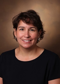 PI: Vicky Morgan, Professor of Radiology and Radiological Sciences, Professor of Biomedical Engineering, Professor of Neurology, Professor of Neurological Surgery
The Morgan Engineering and Imaging in Epilepsy Lab works closely with the departments of Neurology and Neurosurgery to develop Magnetic Resonance Imaging (MRI) methods to improve neurosurgical outcomes, particularly for patients with epilepsy. We directly support clinical care by developing and providing functional MRI to localize eloquent cortex in the brain to aid in surgical planning to minimize functional and cognitive deficits post surgery. Our research focuses on mapping functional and structural brain networks in epilepsy before and after surgical treatment. Ultimately, we aim to use MRI to fully characterize the spatial and temporal impacts of seizures across the brain to optimize management of epilepsy patients. The Morgan lab has on-going research collaborations with the BIEN (Englot) Lab, the Medical Imaging Processing Laboratory (Dawant), the MASI Lab (Landman) and researchers throughout the Vanderbilt Institute of Imaging Science (VUIIS). Contact Vicky Morgan
Neuroimaging and Brain Dynamics (NEURDY) Laboratory
 PI: Catie Chang, Sally and Dave Hopkins Faculty Fellow, Assistant Professor of Computer Science, Assistant Professor of Electrical and Computer Engineering
The goal of our research is to advance understanding of brain function in health and disease. We develop approaches for studying human brain activity by integrating functional neuroimaging (fMRI, EEG) and computational analysis techniques. In one avenue, we are examining the dynamics of large-scale brain networks and translating this information into novel fMRI biomarkers. To enable clearer inferences about brain function with fMRI, we also work toward resolving the complex neural and physiological underpinnings of fMRI signals. Our research is highly interdisciplinary and collaborative, bridging fields such as engineering, computer science, neuroscience, psychology, and medicine. Contact Catie Chang NEURDY Lab
View NEURDY lab video ![]()
Science and Technology for Robotics in Medicine (STORM) Laboratory

Director STORM Lab USA and PI: Keith L. Obstein, Division of Gastroenterology, Hepatology, and Nutrition; Department of Mechanical Engineering—Vanderbilt University
Director STORM Lab UK and PI: Pietro Valdastri, School of Electronic and Electrical Engineering—University of Leeds; Department of Mechanical and Electrical Engineering, Divisions of Gastroenterology, Hepatology and Nutrition—Vanderbilt University
At the STORM Lab we strive to improve the quality of life for people undergoing endoscopy and abdominal surgery by creating miniature and non-invasive capsule robots.
The continuous quest for miniaturization has made the science fiction vision of miniature capsule robots working inside the human body a reality. At the STORM Lab, we are designing and creating mechatronic and self-contained devices to be used inside specific districts of the human body to detect and cure diseases in a non-invasive and minimally invasive manner.
Capsule robots represent a challenging paradigm for both research and learning. They embed sensors, actuators, digital intelligence, miniature mechanisms, communication systems, and power supply, all in a very small volume. Capsule robots may be autonomous or teleoperated, they can work alone or as a team, and they can be customized to fulfill specific functions. We are currently applying capsule robot technologies to early detection and treatment of gastrointestinal cancers (i.e. colorectal cancer, gastric cancer) and are developing a new generation of surgical robots that can enter the patient’s abdomen by a single tiny incision. Building upon these competences, we are always ready to face new challenges by modifying our capsule robots to emerging medical needs. Contact Keith Obstein
View STORM Lab video ![]()
Visual Informatics and Engineering (VINE) Laboratory
 PI: Daniel Moyer, Assistant Professor, Computer Science
Professor Moyer’s group is working to bridge the gap between Machine Learning, and Medical Imaging. We work directly with clinicians and researchers to translate advances in computer vision to better outcomes for patients, and new discoveries in imaging-based scientific fields. While we’re most used to MRI and CT, we’re not afraid to look into new domains, and we’re always happy to meet with new potential collaborators to discuss what might be possible. Contact Daniel Moyer


