Past Symposia
Annual Surgery, Intervention, and Engineering Symposium
2012, 2013, 2014, 2015, 2016, 2017, 2018, 2019, 2020, 2021 , 2022
2022
12th Annual Surgery, Intervention, and Engineering Symposium
Keynote Speaker
Aydogan Ozcan, Ph.D.
Chancellor’s Professor at UCLA
Volgenau Chair for Engineering Innovation
Electrical & Computer Engineering, Bioengineering
HHMI Professor
Howard Hughes Medical Institute
Associate Director
California NanoSystems Institute (CNSI)
Fellow, National Academy of Inventors
Fellow, Guggenheim Foundation
Fellow, AAAS, SPIE, OSA, IEEE, AIMBE, RSC, APS
Date: Wednesday, December 13, 2023
Title:
Virtual Staining of Label-free Tissue Using Deep Learning
Abstract:
Deep learning techniques create new opportunities to revolutionize tissue staining methods by digitally generating histological stains using trained neural networks, providing rapid, cost-effective, accurate and environmentally friendly alternatives to standard chemical staining methods. These deep learning-based virtual staining techniques can successfully generate different types of histological stains, including immunohistochemical stains, from label-free microscopic images of unstained samples by using, e.g., autofluorescence microscopy, quantitative phase imaging (QPI) and reflectance confocal microscopy. Similar approaches were also demonstrated for transforming images of an already stained tissue sample into another type of stain, performing virtual stain-to-stain transformations. In this presentation, I will provide an overview of our recent work on the use of deep neural networks for label-free tissue staining, also covering their biomedical applications
Short Bio:
Dr. Aydogan Ozcan is the Chancellor’s Professor and the Volgenau Chair for Engineering Innovation at UCLA and an HHMI Professor with the Howard Hughes Medical Institute. He is also the Associate Director of the California NanoSystems Institute. Dr. Ozcan is elected Fellow of the National Academy of Inventors (NAI) and holds >60 issued/granted patents in microscopy, holography, computational imaging, sensing, mobile diagnostics, nonlinear optics and fiber-optics, and is also the author of one book and the co-author of >1000 peer-reviewed publications in leading scientific journals/conferences. Dr. Ozcan received major awards, including the Presidential Early Career Award for Scientists and Engineers (PECASE), International Commission for Optics ICO Prize, Dennis Gabor Award (SPIE), Joseph Fraunhofer Award & Robert M. Burley Prize (Optica), SPIE Biophotonics Technology Innovator Award, Rahmi Koc Science Medal, SPIE Early Career Achievement Award, Army Young Investigator Award, NSF CAREER Award, NIH Director’s New Innovator Award, Navy Young Investigator Award, IEEE Photonics Society Young Investigator Award and Distinguished Lecturer Award, National Geographic Emerging Explorer Award, National Academy of Engineering The Grainger Foundation Frontiers of Engineering Award and MIT’s TR35 Award for his seminal contributions to computational imaging, sensing and diagnostics. Dr. Ozcan is elected Fellow of Optica, AAAS, SPIE, IEEE, AIMBE, RSC, APS and the Guggenheim Foundation, and is a Lifetime Fellow Member of Optica, NAI, AAAS, and SPIE. Dr. Ozcan is also listed as a Highly Cited Researcher by Web of Science, Clarivate.
11th Annual Surgery, Intervention, and Engineering Symposium
Keynote:
Martha Morrell MD
Clinical Professor of Neurology, Stanford University
Chief Medical Officer, NeuroPace
Date: Wednesday, December 7, 2022
Title:
Crossing the academia-industry bridge: Delivering a brain-responsive neurostimulator for intractable epilepsy
Abstract:
Relief of suffering from illness or disability is a primary motivation for many who have entered the broad field of medical/biomedical science. This may take the form of fundamental scientific discovery, direct patient care, or of discovery, development and delivery of new therapies that bring benefit. In some instances, therapies provide incremental benefit and less often, true innovation. The efforts to create better treatments for patients in need requires multi-disciplinary and multi-institutional collaboration. If the worlds of academia and industry are seen as separate in intent and motivation, and if these each views the other with distrust, then new therapies and solutions are at risk into falling in the “Valley of Death” between discovery and deliver. Creating obstacles rather than bridges between the 2 entities may deprive persons of therapies that offer relief.
The importance of collaboration between academia and industry will be discussed by sharing the story of a start-up company formed to develop of a responsive brain neurostimulator for persons with drug resistant epilepsy. The story is one of discovery, development and delivery of a novel therapy for a group of patients with a great clinical need. Emphasis will be given to the importance of a team with diverse expertise, the unexpectedly long path filled with accomplishments and disappointments, and the steps that led to eventual success.
Biography:
Dr. Morrell has been Chief Medical Officer of NeuroPace, Inc. and a Clinical Professor of Neurology at Stanford University since July 2004. Before joining NeuroPace, she was the Caitlin Tynan Doyle Professor of Clinical Neurology at Columbia University and Director of the Columbia Comprehensive Epilepsy Center at New York Presbyterian Hospital in New York City. Previously she was on the faculty of the Stanford University School of Medicine where she served as Director of the Stanford Comprehensive Epilepsy Center. A graduate of Stanford Medical School, she completed residency training in Neurology at the University of Pennsylvania, as well as fellowship training in EEG and epilepsy.
Dr. Morrell has been actively involved in helping to bring new therapies to patients. Her responsibilities at NeuroPace include all clinical and pre-clinical research for a novel responsive neurostimulator for the treatment of medically uncontrolled epilepsy. She has been actively involved in investigational trials of new epilepsy therapies as an academic investigator, and has authored or coauthored more than 150 publications.
Service to professional societies includes member of the Board of Directors of the American Epilepsy Society, member and Chair of the Board of the Epilepsy Foundation, member of the Council of the American Neurological Association and Chair of the Epilepsy Section of the American Academy of Neurology. She is an elected Ambassador for Epilepsy of the International League Against Epilepsy and received the American Epilepsy Society’s 2007 Service Award for outstanding leadership and service. She is the immediate past-Chair of the American Society for Experimental Neurotherapeutics.
10th Annual Surgery, Intervention, and Engineering Symposium
Date: December 15, 2021
Keynote:
Natalia Trayanova, PhD, FHRS, FAHA
Murray B. Sachs Endowed Chair
Professor of Biomedical Engineering and Medicine
Director, Alliance for Cardiovascular Diagnostic and Treatment Innovation (ADVANCE)
Johns Hopkins University

Title:
AI-Powered Computational Cardiology
Abstract:
Simulation-driven engineering has put rockets in space, airplanes in the sky, and self-driving cars on the road. Computational approaches, however, have rarely been applied to human health, and trial-and-error is often used to decide on interventions for patients with life-threatening diseases. Working to change this, we have developed personalized imaging-based heart “digital twins”, which digitally replicate the patient’s heart electrical function. We also use machine learning approaches to incorporate additional clinical data in the applications of the heart digital twin technology.
The heart digital twins are models based on patient’s heart scans and clinical data, and are used to simulate the individual patient’s irregular heart rhythm — a major cause of patient morbidity and mortality. We apply the digital twin technology to diagnose which patients with heart disease are likely to develop arrhythmias and thus are at risk of dying suddenly. A digital heart twin is capable of predicting adverse cardiac events as it simulates the effect of potential stressors. Using such a tool, the patient’s risk of sudden cardiac death can be assessed, and a precise decision can be made regarding the benefit-vs-complications of implanting a defibrillator device that can shock the patient back into normal heart rhythm.
We also use the personalized heart digital technology to develop precise treatment for patients suffering from arrhythmias. For each patient, my team determines the location of perpetrator tissue in the heart, so that it can be destroyed by ablation, restoring the heart’s normal rhythm. A mock of the ablation is performed to ensure arrhythmias will not re-emerge post-procedure. This approach not only eliminates the process of trial-and-error in treating heart rhythm disorders but also prevents repeat procedures, which are common in arrhythmia management.
Biography:
Dr. Trayanova is the Murray B Sachs Professor in the Department of Biomedical Engineering at Johns Hopkins University and a Professor of Medicine at the Johns Hopkins School of Medicine. She directs the Alliance for Cardiovascular Diagnostic and Treatment Innovation, a research institute aimed at applying predictive data-driven approaches, computational modeling, and innovations in cardiac imaging to the diagnosis and treatment of cardiovascular disease. She also directs the Computational Cardiology Laboratory.
Using a personalized simulation and machine learning approaches, Dr. Trayanova has developed new methods for predicting risk of cardiac arrest and improving the accuracy of atrial and ventricular catheter ablation therapies. Through her first-of-their-kind personalized virtual hearts, she is pioneering advances in personalized medicine for patients with cardiovascular disease, which promise to profoundly influence clinical decision-making and the delivery of patient care. She is currently conducting FDA-approved clinical trial in simulation-driven treatment for cardiac arrhythmias.
Dr. Trayanova has published over 400 scientific papers, many of them in journals of high impact. She has also given over 300 invited talks, keynotes, and plenary lectures. She is the inventor on 45 patents and patent applications. Her work has received world-wide recognition, and she is the recipient of numerous honors and awards. For her groundbreaking work in computational cardiology, in 2013, she received the NIH Director’s Pioneer Award, the most prestigious recognition of innovation in medical research. In 2019, she was inducted in the Women of Technology International Hall of Fame, an honor conferred only on 5 women each year from around the world. Also in 2019, she received the Distinguished Scientist Award from Heart Rhythm Society. In 2020, she was elected Fellow of the National Academy of Inventors, and received the Douglas Zipes Lectureship Award, bestowed by the Heart Rhythm Society.
Trayanova is also a Fellow of: European Society of Cardiology, American Institute for Medical and Biological Engineering; Heart Rhythm Society; American Heart Association; Biomedical Engineering Society; and International Academy of Medical and Biological Engineering. Her work has been featured by a number of news outlets, including a TEDx talk.
Date: December 9, 2020
Visit our Virtual Research Expo.
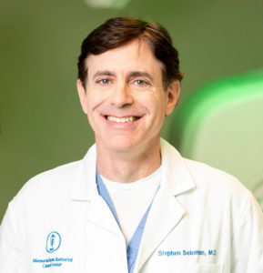
Keynote: Stephen B. Solomon, MD
Enid A. Haupt Chair in Clinical Investigation
Chief of Interventional Radiology
Memorial Sloan Kettering Cancer Center
Professor of Radiology
Weill Cornell Medical College
Title:
Innovations in Interventional Oncology: From Bench to Clinic
Biography:
Stephen Solomon is a physician and scientist driven by innovation, strategic vision, and successful translation to improve clinical care. As Chief of Interventional Radiology over the last 12 years, he has transformed Memorial Sloan Kettering Cancer Center’s (MSKCC’s) Interventional Radiology (IR) Service from 6 faculty and 5,000 annual procedures to over 25 faculty performing 22,000 annual procedures. He has developed an internationally acclaimed Service by recruiting talented and diverse faculty to meet the Center’s academic mission of delivering innovation to the clinical practice, research enterprise, and educational portfolio.
Dr. Solomon himself is regarded as a renowned innovator who holds many patents and has invented and developed several technologies that are being applied in clinical medicine today throughout the world. He has a creative mind that identifies solutions to unmet needs in medicine. He has done this through his own efforts and has instilled this same curiosity and creativity on his team in Interventional Radiology at Memorial Sloan Kettering.
One of his most important contributions to Medicine has been the development of a navigational bronchoscope that has helped spawn the field of Interventional Pulmonary Medicine. Before this invention, bronchoscopy could only visualize structures within the airway. Dr. Solomon developed an electromagnetic position (“GPS”) sensor that tracked the location of the bronchoscope as it moved in the airway. Marrying this location to a CT scan enabled visualization of structures in and out of the airway. This allowed biopsy and other interventions to be performed on structures outside of the airway and allowed guidance to abnormalities seen on a CT scan. This innovation was developed in the laboratory, evaluated in animal models, was translated into clinical trials, and is now a standard tool throughout the world.
Dr. Solomon similarly used this electromagnetic position sensor to track other devices in the body. Another significant success was applying this sensor to catheters in the heart that would track and treat electrical rhythm abnormalities. By combining CT and MR imaging of the heart to the electrical and temporal rhythm of the heart beat, Dr. Solomon helped improve the ability to guide therapy in heart beat dysfunction such as atrial fibrillation. This is also a system that is a standard across the world.
Dr. Solomon’s clinical expertise is image-guided interventions and specifically destroying (i.e. ablating) cancers with heat or cold. He is one of the foremost experts in this field and has developed and translated many of these tools into daily clinical practice at MSKCC and around the world. This has enabled destroying cancer with a simple needle rather than having to undergo a major surgical resection. This technology is routinely applied in lung cancers, liver cancers, bone cancers, kidney cancers, and others.
Dr. Solomon has worked with Nobel Laureate Jim Allison to demonstrate how using thermal ablation in mice can create an immune response to the dead ablated tissue and how this immune response can be trained to fight cancer in other parts of the body, creating a “personalized cancer vaccine.” Dr. Solomon has helped translate this work into ongoing clinical studies.
Dr. Solomon’s laboratory has also experimented with a variety of energy sources to destroy cancer. One of the most interesting has been the application of electrical fields on cancer cells. The field of electroporation has been around for a while and has allowed a cell’s membrane to be temporarily permeable. Dr. Solomon has experimented with irreversible electroporation which permanently destroys cells by irreversibly destroying their cell membranes. This has benefits of destroying cells but not the proteins that create the structure of an organ. This may allow preservation of the organ’s function (e.g. bile ducts or bronchi) while still killing the cancer cells. With his engineering partner he developed a new method of killing cancer cells called “e-stress” which through continued cell membrane depolarizations, that the cell must use energy to correct, “exhausts” the cells of its energy supply and leads to cell death.
While CAR T-cells have made a significant impact on “liquid tumors” such as lymphoma, it still has not been very successful in solid tumors. Dr. Solomon has teamed up with colleagues at MSKCC to improve CAR T-cell delivery with image-guidance and to apply electrical fields to enhance CAR T-Cell delivery to solid tumors. This has led to receipt of an NIH R01 grant.
As Precision Medicine has become central to cancer care, so too has the image-guided biopsy to collect the tissue necessary for genetic analysis. One of the challenges of image-guided biopsy is knowing while the patient is in the biopsy suite that the appropriate amount of tissue has been collected. Without knowing this a patient may return for treatment 3 weeks later to be told that insufficient tissue was collected to perform molecular analysis. Dr. Solomon has led development of an optical spectroscopy device that analyzes the tissue at the time of biopsy to determine sufficient quantity for molecular studies.
Dr. Solomon has been instrumental in a number of additional advances in cancer care ranging from molecular-guided PET interventions to protective displacement of organs undergoing radiation therapy to intra-arterial delivery of chemotherapy for lung metastases. He has encouraged his team to think creatively and apply the tools of imaging to solve the unmet challenges in medicine. This has led the MSKCC IR team to be one of the most academically prolific and well-funded worldwide with research across multiple DMT disease states.
Dr. Solomon has over 250 peer-reviewed publications, chapters, and reviews. He is a graduate of Harvard College and Yale School of Medicine. He completed his radiology residency at Johns Hopkins and his fellowship at New York Presbyterian Hospital/Weill Cornell Medical Center. He was a faculty member at Johns Hopkins for 7 years before joining MSKCC as the Director of The Center for Image Guided Intervention and then Chief of Service. He has recently been granted the Enid A. Haupt Endowed Chair in Clinical Investigation at Memorial Sloan Kettering.
Date: Wednesday, December 11, 2019
View the program here
Keynote: Louis Kavoussi, MD
Chairman of Urology for Northwell Health
and
Waldbaum Gardner Professor of Donald and Barbara Zucker School of Medicine at Hofstra/Northwell
Title: “End Game” A surgeon’s perspective on the future of technology and innovation in healthcare.
Abstract:
Surgery can be used to treat many pathological conditions; however, the price of cure includes potential convalescence, discomfort, impact on cosmesis, variable functional outcomes and complications. Improvements in techniques as well as technology have decreased morbidity and mortality once commonly associated with an operation. Currently there is still inconsistency in outcomes related to innate differences among surgeons as well as patients. Advances in technology will allow surgery to evolve into the ideal patients’ desire. A guaranteed perfect outcome absent of problems and disruption of individual normality. We rely upon our engineering colleagues to help deliver this reality.
Biography:
Dr. Kavoussi completed his undergraduate degree at Columbia University and medical degree at the State University of New York at Buffalo. He obtained his urologic training at Washington University of St. Louis and directly following residency was named Chief of Urology at the Jewish Hospital of St. Louis. In 1991 he was appointed Assistant Professor at Harvard School of Medicine and Director of Endourology at the Brigham and Women’s Hospital. In 1993 he joined the faculty of Johns Hopkins University School of Medicine where he was Vice Chairman of Urology and Patrick C. Walsh Distinguished Professor. Dr. Kavoussi is currently the Chair of Urology for the Northwell Health (Formerly North Shore-LIJ Health System) and the Waldbaum-Gardner Distinguished Professor of Urology at the Donald and Barbara Zucker School of Medicine at Hofstra/Northwell.
Dr. Kavoussi has made many important contributions to urology.
He was part of several first teams that engendered many of the minimally invasive approaches we use today including the laparoscopic nephrectomy, laparoscopic donor nephrectomy and laparoscopic prostatectomy. His contributions have been documented in over 400 peer reviewed publications. He has edited multiple texts including his role as a co-editor of Campbell-Walsh Urology, Smith’s Endourology, Atlas of Retroperitoneal Surgery and Handbook of Surgical Techniques.
He helped found the first robotics laboratory dedicated to urological applications and developed telerounding and teleinteraction with patients for which he was a Smithsonian Computerworld Medalist. His contributions to the field of urology earned him the Golden Cystoscope Award of the American Urological Association, The Lifetime Achievement Award from the American Kidney Foundation and the 2018 Distinguished Contribution Award from the American Urological Association.
Dr. Kavoussi has mentored over 35 fellows in minimally invasive surgery who are academic leaders around the world. This achievement was recognized through him being awarded the Endourology Society Mentoring Award in 2014. He has served the field of urology in many areas. He has been on the Board of the Endourology Society. He currently is treasurer of the New York Section of the American Urological Association (in line for Presidency coming year). He is also President elect of the Society for Academic Urologists.
Date: December 12, 2018
View program by clicking here.
Keynote:
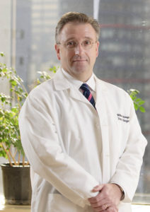
William R. Jarnagin, MD,
Attending Surgeon
Leslie H. Blumgart, M.D. Chair in Surgery
Chief, Hepatopancreatobiliary Service
Memorial Sloan-Kettering Cancer Center
Professor of Surgery
Cornell University Weill College of Medicine
Title:
Innovations in Surgical Data Science with Oncologic Application
Abstract:
Intrahepatic cholangiocarcinoma (IHC) is a devastating, largely incurable cancer, with a rising world-wide incidence and few effective treatment options. Systemic chemotherapy is the mainstay of treatment but is limited. Advances in genomics and radiomics are starting to improve our ability to classify tumors, stratify risk and tailor treatment. We are developing imaging tools to inform the care, treatment, and cure of cancer patients. We will discuss our recent work to optimally select patients for therapy, elucidate mechanisms of cancer progression, identify high-risk patients, and guide surgical resection with imaging technology.
Biography:
Dr. William R. Jarnagin was raised outside of Boston, Massachusetts and earned his undergraduate degree in chemistry from Dartmouth College in 1982, a Master’s degree in chemistry from Brandeis University in 1984 and an MD from Rush Medical College in 1988. He completed his training in general surgery at the University of California, San Francisco in 1996. From 1990-93, he completed a research fellowship at the Liver Center Laboratory at San Francisco General Hospital. From 1996-97, he served as the Hepatobiliary Fellow at Memorial Sloan-Kettering Cancer Center (MSKCC). Since 1997, he has been an attending surgeon at Memorial Sloan-Kettering Cancer Center, where he has served as Chief of the Hepatopancreatobiliary Service since 2008 and was a Vice-Chairman of the Department of Surgery from 2006-2010. He holds the Leslie H. Blumgart Chair in Surgery and is Professor of Surgery at Weill Medical College of Cornell University.
Dr. Jarnagin’s research has focused on genomics, novel therapies and biomarkers of treatment response in patients with biliary tract cancer, intraoperative navigation systems, and improvements in intraoperative management during major liver and pancreas resection. He has authored or co-authored 400 articles in peer-reviewed journals, over 75 book chapters or invited reviews and has co-edited four textbooks. He has served as the HPB Section Editor for Annals of Surgical Oncology and is a member of the editorial boards of the Journal of the American College of Surgeons, HPB and the European Journal of Surgical Research. In addition to the IHPBA and AHPBA, he is a member of several surgical societies, including SUS and ASA. He was a member of the AHPBA Executive Council for over 10 years, serving as the Program Chair (2007-08), Treasurer (2009-11), President (2011-2012), and he currently serves as Treasurer of the IHPBA.
Invited Lecture #1
Jin U. Kang, PhD,
Jacob Suter Jammer Professor (primary)
Department of Electrical and computer engineering
The Johns Hopkins university
and
Professor (Secondary)
Department of Dermatology
Johns Hopkins Medicine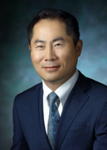
Title:
Image-Guided Advanced Surgical Systems and Techniques for Microsurgery
Abstract: The advances in 3D optical imaging and sensing technologies are enabling the development of the next generation of smart surgical devices and systems. In this intelligent “smart” surgical platform, optical sensors/imagers, robotics, and computers are combined with surgical devices and systems to attain surgical outcomes beyond free-hand human capabilities. In our laboratories, we have been developing real-time intraoperative optical coherence tomography systems specifically for the development of practical and smart microsurgical tools and 3-D image guided surgical systems that enhance the surgeon’s ability to visualize optically transparent tissues, to identify and track visually transparent tissue edges and tools, to maintain safe surgical positions, to detect early instrument contact with tissue and to assess depth of instrument penetration into tissues. These innovations enhance the surgeon’s ability to achieve surgical objectives, diminish surgical risk, and improve outcomes. In this talk, I will summarize our recents efforts in optical image-guided robotic surgical procedures and future directions.
Biography:
Jin U. Kang, Jacob Suter Jammer Professor of Electrical and Computer Engineering, is an expert in optical imaging, sensing, fiber optic devices and photonic systems. He holds a joint appointment in the Department of Dermatology at the Johns Hopkins University School of Medicine and is a member of the Johns Hopkins’ Kavli Neuroscience Discovery Institute and the Laboratory for Computational Sensing and Robotics. Kang chaired the Department of Electrical and Computer Engineering from 2008-2014.
Kang’s research is focused on developing novel optical techniques and devices for a wide range of biomedical applications. Kang has been developing endoscopic common-path fiber optic optical coherence tomography (OCT) techniques for medical imaging and sensing; these systems have enabled the development of microsurgical and robotic tools that allow safer, more precise surgical outcomes. Kang’s group was the first to implement and demonstrate real-time 4D OCT systems that could allow surgeons to monitor surgical sites in 3-D video during surgery.
Kang has also been working to create a “smart” tool system to help surgeons more accurately and safely perform microsurgeries in areas like retinal surgeries, cornea transplants, vascular surgeries and cochlear implants. He recently launched a JHU Fast Forward startup company, LIV (Live Imaging Vision) Med Tech Inc., to work on these image-guided robotic tools. Other projects involve building real-time Doppler 3-D imaging systems for intraoperative assessment of surgeries like carotid endarterectomy and cerebrovascular surgery.
A holder of over 20 patents and 176 refereed journal publications, Kang is a program chair of CLEO A&T and a fellow of the Optical Society of America, the International Society for Optics and Photonics (SPIE), and the American Institute for Medical and Biological Engineering (AIMBE). He is a recipient of the ONR Young Investigator Award, the Australian Institute of Advanced Studies Fellowship, a NASA Faculty Fellowship, the Oak Ridge Institute of Science and Education fellowship, and the Brain Korea Distinguished Faculty Fellowship.
Kang was a topical editor of Optics Letters and is an editorial board member of the Journal of the Optical Society of Korea and Chinese Optics Letters.
Kang received his B.S. degree in physics from Western Washington University in 1992, and a master’s degree and Ph.D. in optical science and electrical engineering from the University of Central Florida in 1993 and 1996, respectively. He worked as a research engineer for the U.S. Naval Research Laboratory in Washington, D.C., prior to joining the faculty of Whiting School of Engineering in 1998.
Invited Lecture #2
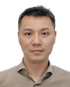
S. Kevin Zhou, PhD
Professor, Institute of Computing Technology,
Chinese Academy of Sciences
Title:
Machine learning + knowledge modeling: medical image recognition, segmentation, parsing
Abstract:
The “Machine learning + Knowledge modeling” approaches, which combine machine learning with domain knowledge, enable us to achieve start-of-the-art performances for many tasks of medical image recognition, segmentation and parsing. In this talk, we present real success stories of such approaches and proceed to elaborate deep learning, the most powerful machine learning method. We demonstrate that an extra performance boost is rendered when deep learning is combined with knowledge modeling.
Biography:
Dr. S. Kevin Zhou is currently a professor at Institute of Computing Technology, Chinese Academy of Sciences. He was a Principal Key Expert of Image Analysis at Siemens Healthcare Technology. His research interests lie in computer vision and machine learning and their applications to medical image recognition and parsing, face recognition and modeling, etc. Dr. Zhou has published over 150 book chapters and peer-reviewed journal and conference papers, has registered over 250 patents and inventions, has written two research monographs, and has edited three books. His two most recent books are entitled “Medical Image Recognition, Segmentation and Parsing: Machine Learning and Multiple Object Approaches, SK Zhou (Ed.)” and “Deep Learning for Medical Image Analysis, SK Zhou, H Greenspan, DG Shen (Eds.).” He has won multiple awards honoring his publications, patents and products, including Thomas Alva Edison Patent Award (2013), R&D 100 Award or Oscar of Invention (2014), Siemens Inventor of the Year (2014), and UMD ECE Distinguished Aluminum Award (2017). He has been an associate editor for IEEE Trans Medical Imaging and Medical Image Analysis journals,an area chair for CVPR and MICCAI, and elected as a fellow of American Institute of Biological and Medical Engineering (AIMBE).
Invited Lecture #3
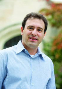
Gregory S. Fischer, PhD
William Smith Dean’s Professor, Mechanical Engineering & Robotics Engineering
Director, Worcester Polytechnic Institute
Title:
Image-Guided Robotic Surgery: In-situ MRI Guidance for Enhancing Robot-Assisted Cancer Therapy
Abstract:
Interactively updated intraoperative medical imaging affords the opportunity to monitor and guide interventional procedures. The real-time feedback enables “closed loop medicine” wherein we ensure that the treatment plan is implemented as intended. In order to take the most advantage of robots in surgery, we work towards integrating real-time medical imaging with the interventional procedure to provide as much information to a surgeon during a procedure as possible and using that information in a way to produce better outcomes. MRI can offer high resolution 3D imaging with high soft tissue contrast, multi-modality imaging for tumor localization, thermal monitoring, and interactively updated speed, making it ideal for monitoring and guiding interventions. However, challenges with the high magnetic field, time varying magnetic gradient, strong RF signals, and high sensitivity to RF noise make leveraging these capabilities a challenge. We have developed a modular approach to MRI-compatible robotics including the software, control hardware, and mechanical systems, and have used this approach to develop robotic systems for image-guided diagnosis and therapy of prostate cancer and for stereotactic neurosurgical interventions where we can perform surgical manipulation under live MR imaging. A system for percutaneous access to the prostate has been used in a 30-patient trial for prostate cancer biopsy at the Brigham and Women’s Hospital in Boston, MA. And, an MRI-compatible stereotactic neurosurgery robot intended for conformal thermal ablation utilizing interstitial therapeutic ultrasound is in preclinical trials at UMass Medical School in Worcester, MA. This robot combines precision alignment of the probe based on intraoperative imaging that can account for brain shift, monitoring of the probe insertion, and live thermal monitoring of the dose delivered. Current work funded under the NIH NCI Academic-Industry Partnership program is focused on utilizing real-time feedback coupled with interactive robotic control of the ablation probe to produce precise conformal boundaries for ablation of irregularly shape Glioblastomas. Other work in the WPI Automation and Interventional Medicine (AIM) Robotics Research Laboratory includes task automation and intelligent teleoperation of the daVinci surgical robot, soft wearable assistive robots, and socially assistive robots for augmenting Autism therapy.
Biography:
Dr. Gregory Fischer is the William Smith Dean’s Professor of Mechanical Engineering & Robotics Engineering with an appointment in Biomedical Engineering at Worcester Polytechnic Institute. He received his PhD from Johns Hopkins University in Mechanical Engineering and an MS in Electrical Engineering where he was a member of the NSF Engineering Research Center for Computer Integrated Surgery (ERC-CISST). He is the Director of PracticePoint at WPI (http://practicepoint.org), a newly launched MassTech-supported research, development, and commercialization alliance centered built to advance healthcare technologies and launch new smart and secure medical cyber-physical systems. PracticePoint comprises integrated advanced manufacturing capabilities co-located with point of practice clinical care settings, and is poised to advance healthcare in: Medical Cyber-physical Systems, Surgical Robots, Medical Imaging and Image-Guidance, Sensing, Actuation, and Control of Medical Devices, Wearable robotics, Advanced manufacturing for personalized healthcare, Cybersecurity for medical devices, Data analytics for clinical decision support. Dr. Fischer also leads the WPI Automation and Interventional Medicine Robotics Research Laboratory (AIM Lab: http://aimlab.wpi.edu) with research focus including: an MRI-compatible robotic system for image-guided conformal ablation of deep brain tumors currently in pre-clinical trials, a robotic system for precision targeted MRI-guided prostate cancer biopsy currently in clinical trials, wearable soft robotic devices for rehabilitation and assistance with activities of daily living, and social robots for providing therapy.
Contact:
Gregory S. Fischer, PhD
Wednesday, December 13th, 2017
View the symposium program by clicking here.
keynote delivered by
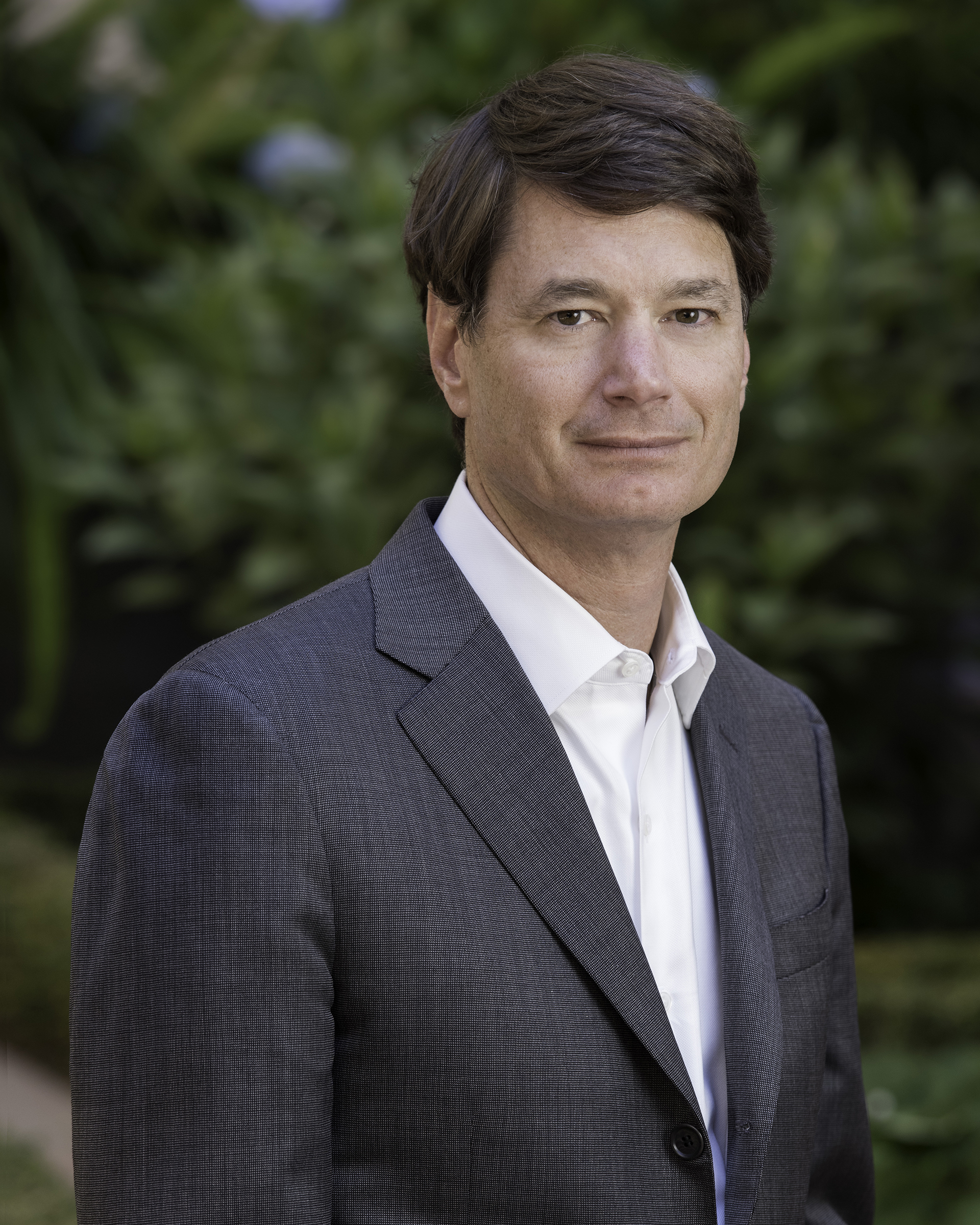
Eben Rosenthal, MD
Ann & John Doerr Medical Director
Stanford Cancer Center
Title:
“Leveraging Light in the OR: Finding an Optical Contrast Agent”
Abstract:
Over the past two decades, synergistic innovations in imaging technology have resulted in a revolution in which a range of biomedical applications are now benefiting from fluorescence imaging. Specifically, advances in fluorophore chemistry and imaging hardware, and the identification of targetable biomarkers have now positioned intraoperative fluorescence as a highly specific real-time detection modality for surgeons in oncology. In particular, the deeper tissue penetration and limited autofluorescence of near-infrared (NIR) fluorescence imaging improves the translational potential of this modality over visible-light fluorescence imaging. Rapid developments in fluorophores with improved characteristics, detection instrumentation, and targeting strategies led to the clinical testing in the early 2010s of the first targeted NIR fluorophores for intraoperative cancer detection. The foundations for the advances that underline this technology continue to be nurtured by the multidisciplinary collaboration of chemists, biologists, engineers, and clinicians. In this Review, we highlight the latest developments in NIR fluorophores, cancer-targeting strategies, and detection instrumentation for intraoperative cancer detection, and consider the unique challenges associated with their effective application in clinical settings.
Bio:
Eben Rosenthal is a surgeon-scientist who serves as the John and Ann Doerr Medical Director of the Stanford Cancer Center. He works collaboratively with the Stanford Cancer Institute and Stanford Health Care leaders to set the strategy for the clinical delivery of cancer care across Stanford Medicine and growing cancer networks. He is a professor of Otolaryngology and Radiology (courtesy) and a member of Molecular Imaging Program at Stanford (MIPS). He continues to be clinically active as an oncologic and microvascular reconstructive surgeon.
Dr. Rosenthal has conducted multiple early phase clinical trials for diagnostic and therapeutic agents for the treatment of solid tumors. He has been NIH funded for over a decade in targeted, translational research. He is part of a multidisciplinary team of clinical and basic scientists that have successfully performed preclinical studies, nonhuman primate IND-enabling studies, successfully performed the first in human clinical trials using fluorescently labeled antibodies as a cancer specific contrast agent for use in the operating room. Ongoing clinical trials include brain, pancreas, skin and head and neck tumors.
2016
Wednesday, December 14th, 2016
keynote delivered by
Christopher P. Austin, MD,
Director, National Center for Advancing Translational Science
at the National Institutes of Health
 Catalyzing Translational Innovation
Vanderbilt University Light Hall
Abstract:
The process by which observations in the laboratory or the clinic are transformed into demonstrably useful interventions that tangibly improve human health is frequently termed “translation.” This multi-stage and multifaceted process is poorly understood scientifically, and the current research ecosystem is operationally not well suited to the distinct needs of translation. As a result, biomedical science is in an era of unprecedented accomplishment without a concomitant improvement in meaningful health outcomes, and this is creating pressures that extend from the scientific to the societal and political. To meet the opportunities and needs in translational science, NCATS was created as NIH’s newest component in December 2011, via a concatenation of extant NIH programs previously resident in other components of NIH. NCATS is scientifically and organizationally different from other NIH Institutes and Centers. It focuses on what is common to diseases and the translational process, and acts a catalyst to bring together the collaborative teams necessary to develop new technologies and paradigms to improve the efficiency and effectiveness of the translational process, from target validation through intervention development to demonstration of public health impact. This talk will provide an overview of NCATS mission, programs, and deliverables, with a view toward future developments.
Christopher P. Austin, M.D.
Director, National Center for Advancing Translational Sciences, National Institutes of Health
Christopher Austin is director of the National Center for Advancing Translational Sciences (NCATS) at the National Institutes of Health (NIH). NCATS’ mission is to enhance the development, testing and implementation of diagnostics and therapeutics across a wide range of human diseases and conditions. The Center collaborates with other government agencies, industry, academia and the nonprofit community. Before joining NIH in 2002, Austin directed research and drug development programs at Merck, with a focus on schizophrenia. His earned his M.D. from Harvard Medical School, and completed clinical training at Massachusetts General Hospital and a research fellowship in genetics at Harvard.
View the symposium program by clicking here.
2015
Wednesday, December 16, 2015
keynote delivered by
Elizabeth C. Tyler-Kabara, M.D., Ph.D.
Associate Professor of Neurological Surgery and Bioengineering, University of Pittsburg School of Medicine
McGowan Institute for Regenerative Medicine Division of Pediatric Neurosurgery
“From science fiction to real world medical applications”
Abstract:
Science fiction has promised the interface between . humans and machines for decades Television brought us the 6 Million Dollar Man and the movies showed us that Luke Skywalker’s prosthetic hand was fully functional and seamlessly integrated. The medical device industry has brought some of these promises to patients with smart prosthetic devices. Now technology has made interfacing the brain directly to assistive devices a reality. The popular television show Grey’s Anatomy portrayed brain computer interfaces (BCIs) and their ability to move a virtual reality arm or a robotic arm. In fact, BCI’s can be used to control a robotic arm in up to 10 degrees of freedom or bilateral arms in simple tasks. BCI’s can be used to control other assistive and recreational computer programs. There is evidence now that BCIs can provide tactile information from these devices. Patients with quadriplegia due to a variety of disorders have been able to use BCIs to control both virtual reality devices and actual robotic devices. These advances in BCI technology will be the spring board for making this technology a clinical tool.
Biography:
Elizabeth C. Tyler-Kabara, MD, PhD completed her bachelor’s degree at Duke University, double majoring in biomedical and electrical engineering, in 1989. After leaving Duke, she worked at the National Institutes of Health as a biomedical engineer, developing and testing molecular biology software, developing a strategic plan for implementing computer networking, and recruiting a head for the newly formed Computational Biology Group. She left the NIG to attend Vanderbilt University, earning her MD and PhD in 1997. Her graduate research in the Department of Molecular Physiology and Biophysics investigated the neurophysiology of the corticostriatal synapse. This served as the basis for her interest in neuromodulation, which has been a key aspect of her subsequent clinical research activities.
2014
Wednesday, December 10, 2015
keynote delivered by
Robert M. Sweet, M.D., FACS
Associate Professor Urology, William L. Anderson Endowed Chair
Director Medical School Sim Programs,
Director of the Kidney Stone Program
University of Minnesota,
President Society of Laparoendoscopic Surgeons
“The Emergence of Simulation Science for Healthcare”
Abstract:
The science of simulation in healthcare, while in its infancy, is an exciting new discipline that draws upon collaborative expertise involving healthcare societies and providers, industry, patients, engineers and computer scientists graphic artists, sculptors, human factors, and educational researchers. This lecture will go over the rationale and opportunities for health care applications. The portfolio of projects across the Center for Research in Education and Simulation Technologies (CREST) at the Unviversity of Minnesota will be shared and discussed, including the development of a human tissue property database and the creation of an ecosystem of simulation projects including state-of-the-art artificial tissue analogues, next generation manikins, 3-D anatomical virtual models for practitioner and patient-education and full VR procedural trainers.
Biography:
Dr. Rob Sweet received his medical degree (alpha omega alpha) from the University of Minnesota in 1997. After a urology residency at the University of Washington in Seattle in 2003, he became Attending Physician/Acting Assistant Professor of Urology and held a 2-year Health Policy Scholarship from the American Foundation for Urological Diseases (AFUD). In 2004 Dr. Sweet co-founded the Institute for Surgical and Interventional Simulation (ISIS) at the University of Washington. He currently holds the positions of Associate Professor of Urology at the University of Minnesota, and is Director of the Medical School’s Simulation Programs and the Kidney Stone Program. He is the Principal Investigator of numerous simulation research and development projects including the University of Minnesota MedSim Combat Casualty Training Consortium.
2013
Wednesday, December 11, 2013
delivered by
L. Nelson Hopkins, MD, FACS
SUNY Distinguished Professor
Professor of Neurosurgery and Radiology
State University of New York at Buffalo
Director, Toshiba Stroke and Vascular Research Center
CEO of the Jacobs Institute
“Innovations in Vascular Care”

Abstract:
According to the American Heart Association’s 2013 statistics update, more than one in three American adults currently have one or more types of cardiovascular disease. Stroke remains the leading cause of long-term disability and affects 795,000 individuals annually. The prevalence and associated financial burden of cardiovascular and cerebrovascular diseases are projected to increase in the future.
This talk will provide an overview of the importance of a comprehensive vascular center in providing care for a wide range of cardiovascular and cerebrovascular diseases. The origins, goals, and current experience of the Gates Vascular Institute (GVI) as an example of a modern vascular center will be discussed. The GVI structure includes dedicated surgical and minimally invasive interventional labs, a new emergency department, and dedicated CT and MRI scanners. Its collaboration with the University at Buffalo is possible through a dedicated four-floor clinical translation research center, located in the same building. The Jacobs institute is an integral part of the GVI and is the institute’s main hub for medical breakthroughs, innovative treatments, and new economic opportunities.
We will review current standards and designation processes for primary and comprehensive stroke center certification and their role in the management of ischemic and hemorrhagic stroke. We will discuss the importance of quality assurance, the current financial status of health care, and future directions in the management and treatment of vascular disease in the United States.
Biography:
Dr. L. Nelson Hopkins continues working in UB Neurosurgery but recently stepped down as Professor and Chairman of the Department of Neurosurgery at the Department of Neurosurgery at the University at Buffalo from 1989 – 2013. After completing his undergraduate studies at Rutger University, Dr. Hopkins earned a doctor of medicine degree cum laude from Albany Medical College. His post-graduate training included a surgical internship at Case Western Reserve, followed by neurology and neurosurgical training at State University of New York at Buffalo.
Dr. Hopkins has also been active in medical technology entrepreneurship and serves on the board of three medical device start up companies.
2012
keynote delivered by
Dr. Reed Omary, M.D., M.S.
Carol D. & Henry P. Pendergrass Professor
and
Chairman of the Vanderbilt University Department of Radiology and Radiological Sciences
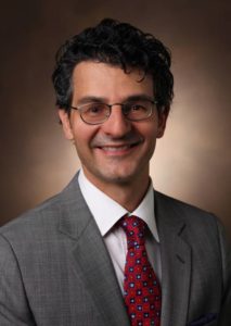
Biography:
Dr. Omary is a translational physician-scientist who is active in education, patient care, research, and administration.
His clinical practice in interventional radiology is focused on image-guided therapies for hepatocellular carcinoma (HCC). Dr. Omary’s major interests include the use of MRI to guide and functionally monitor catheter-based drug delivery. Dr. Omary has clinically translated these techniques using an innovative combined magnetic resonance imaging-x-ray digital subtraction angiography unity.
Dr. Omary is principal investigator (PI) on two separate NIH RO1 grants (one clinical, one pre-clinical) aiming to improve therapies for HCC. He has previously served as PI or co-PI on NIH, RSNA, and SIR training grants. His trainees at the medical student, graduate student, resident, and junior faculty levels have been awarded 32 national and 17 local research prizes. A Fellow in the Society of Interventional Radiology (SIR), Dr. Omary served as Chair of the SIR Foundation Grant Review Study Section from 2007-2011. He is a full member of the NIH Medial Imaging (MEDI) study session, and has participated in numerous other NIH grant review panels.


