Overview
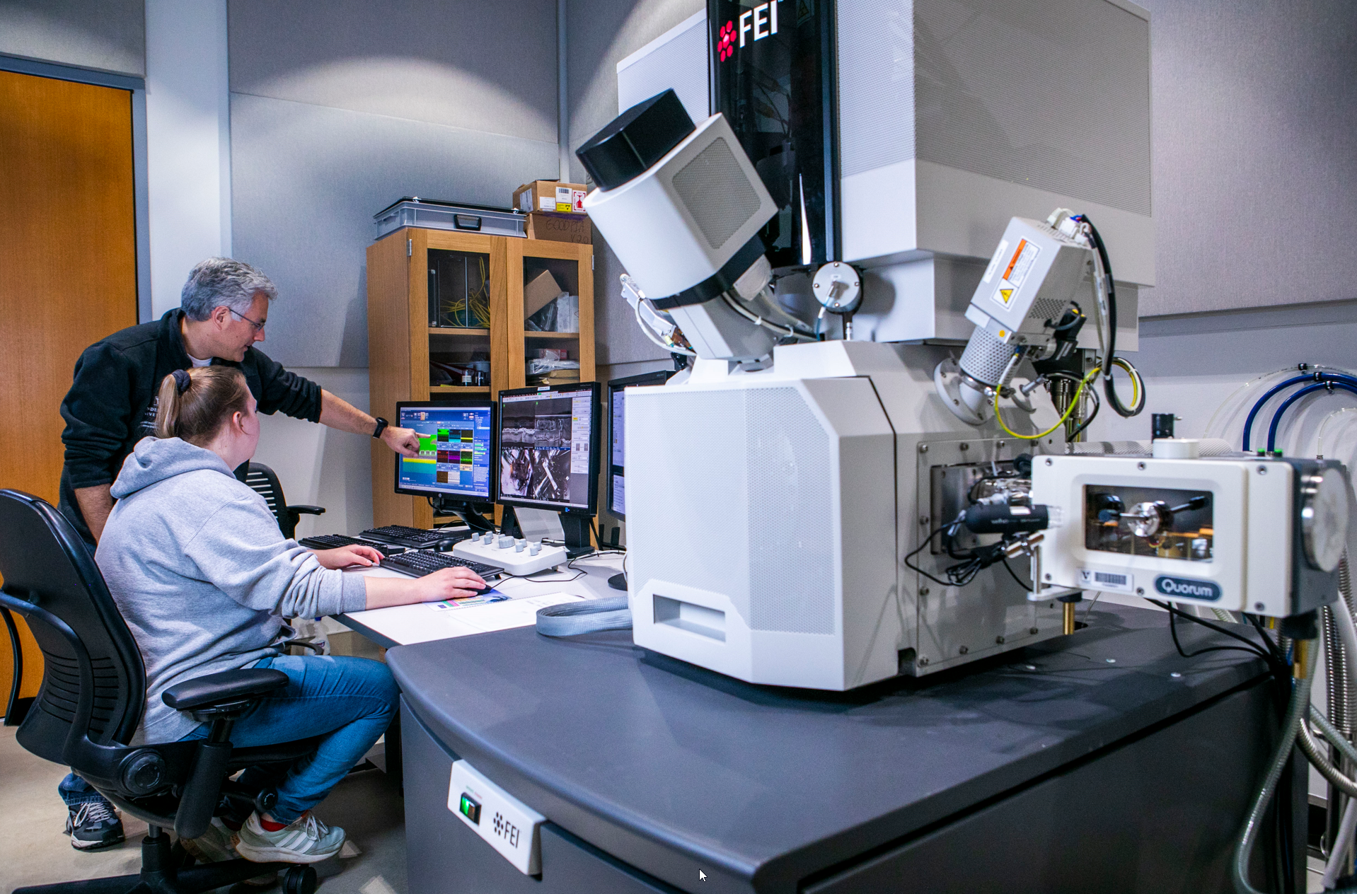
The Helios Nanolab combines the high spatial resolution afforded by an SEM with high precision milling and Pt deposition with a Ga ion beam. With the use of the Easylift nanomanipulator and AutoTEM4 software, thin cross sections of materials can be prepared for high resolution imaging and chemical mapping in the Osiris TEM/STEM. The Nanobuilder software allows users to create complex milling and patterning procedures that can performed un-attended. The addition of the Quorum PP3010 enables cryo-SEM and cryo-FIB-SEM imaging of soft, fully hydrated, beam sensitive materials and biological samples.
Capabilities
- Imaging
- High Resolution SEM Optimized for Low Voltage
- In-Lens Secondary and Backscattered Electron Detector
- Everhart-Thornley Secondary Electron Detector
- Advanced Backscattered Electron Detection (Concentric Backscatter Detector)
- Segmented STEM3 Scanning Transmission Detector
- ICE Secondary Ion and Electron Detector
- Spectroscopy
- Energy Dispersive Spectroscopy (Bruker X-max 50 SDD)
- Milling and Patterning
- Focused Ion Beam Milling and Patterning (4 nm with 30 kV Ga ions)
- Pt E-beam and I-beam Deposition
- GDSII Compatible Nanobuilder Software
- TEM Sample Preparation
- TEM Lift-Out Lamella Preparation (Easylift EX and AutoTEM4 Software)
- Volume FIB-SEM Imaging
- Automated Large Volume Imaging (Auto Slice and View 4)
- Volume Alignment and Segmentation with Amira Software
- Cryo-SEM and Cryo-FIB-SEM
- Quorum PP3010T Cryo Preparation System
- Automated Cryo-ET Sample Preparation (SerialFIB)
- Build Your Own Automation
- Advanced Scripting for Python-based Automation
Applications
- Micro to Nanoscale Imaging and Measuring
- Nanoscale Chemical Mapping and Quantification
- STEM in SEM Imaging for Low Voltage Transmission Imaging
- TEM Sample Preparation from Bulk Samples
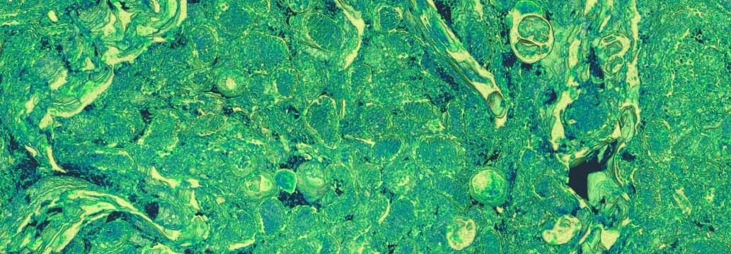
- Volume Imaging of Fixed Tissue or Layered Materials
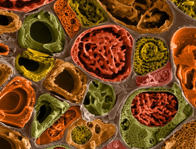
- Cryo-SEM of Hydrated Materials and Biological Samples
- Cryo-ET Sample Preparation
- Nanolithography
- Nanoscale Soldering
Contact
-

Dr. James McBride
VINSE Advanced Imaging
- 019 Engineering and Science Building