Overview
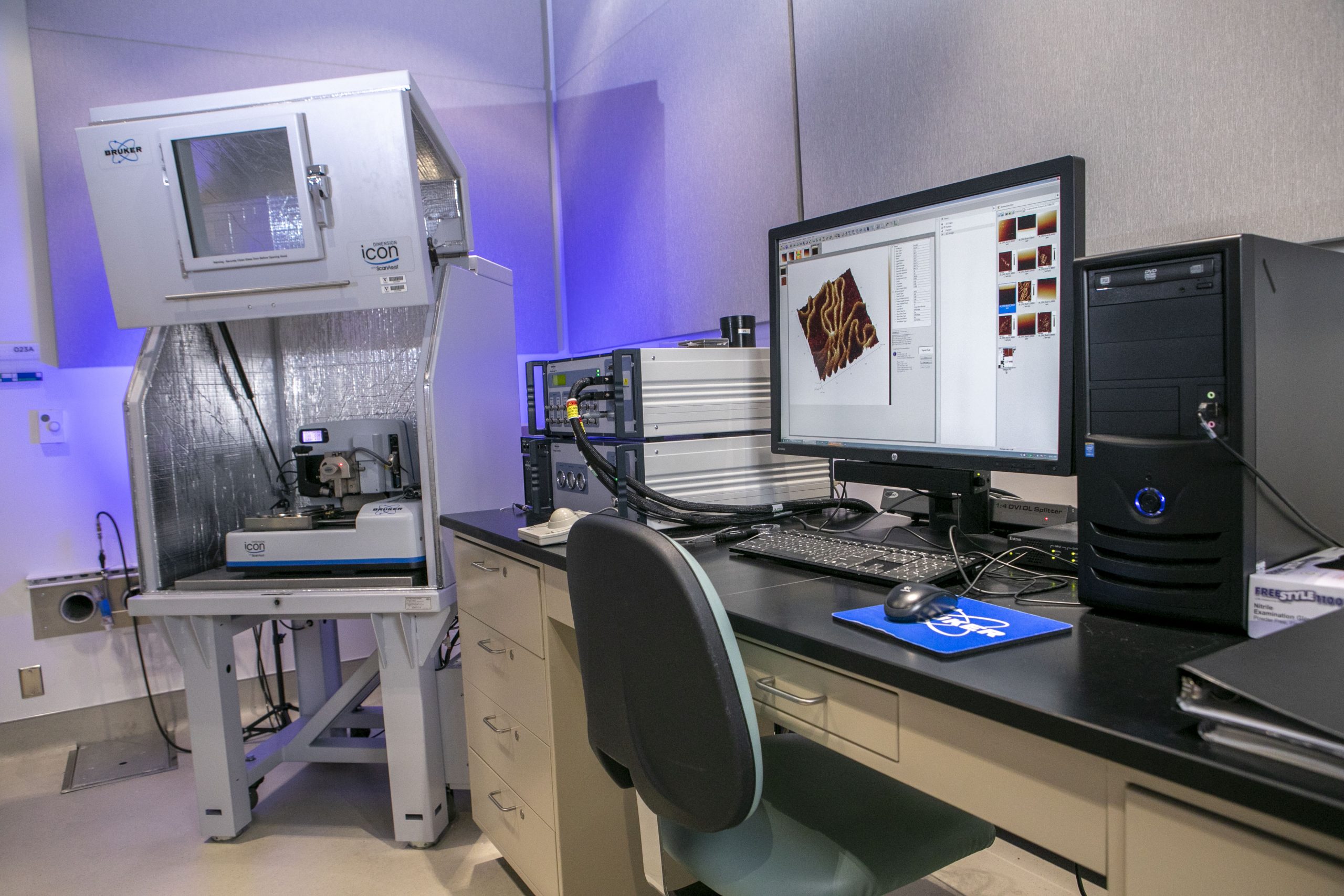
The Dimension Icon® Atomic Force Microscope brings new levels of performance, functionality, and AFM accessibility to nanoscale researchers in science and industry. The culmination of decades of large-sample AFM technology, the system has been designed from top to bottom to deliver revolutionary low drift and low noise that allows users to achieve artifact-free images in minutes instead of hours. The Icon is also equipped with a heater controller to be used in conjunction with the fluid cell kit. The cell kit allows real-time observations of samples under native conditions while providing sample heating and temperature control when liquids are being studied.
Nanomanipulation and nanolithography capabilities integrate object manipulation and surface modification in air and liquid with nanoscale imaging. Both immediate and programmed commands are available to probe a sample between image scans of the field of view. Proprietary ScanAsyst® automatic image optimization technology enables easier, faster, and more consistent results, regardless of user skill level. The Icon’s uncommon ease of use, ultimate performance, exceptional productivity, and superior versatility make it an ideal choice for practically every AFM application.
Capabilities
- X-Y scan range: 90µm x 90µm typical
- Z range: 10μm typical in imaging and force curve modes
- Sample size/holder: 210mm vacuum chuck for samples, less than 210mm, less than 5mm thick
- Vertical noise floor: less than 30pm
- X-Y position noise: less than 0.15nm
- Motorized position stage (X-Y axis): 180mm × 150mm inspectable area
- Microscope optics: 5-megapixel digital camera
- Digital zoom and motorized focus
- Controller NanoScope V
- Vibration isolation: Integrated, pneumatic
- Acoustic isolation
AFM Modes
- Contact Mode
- Tapping Mode
- PhaseImaging
- ScanAsyst/PeakForce mode
- PeakForce QNM
- Force Spectroscopy/Force Curves
- Force Volume++
- Piezo Response Force Microscopy
- Lateral Force Microscopy (LFM)
- Magnetic Force Microscopy (MFM)
- Electric Force Microscopy (EFM)
- Kelvin Probe Force Microscopy (KPFM-AM)/Surface Potential
- Scanning Tunneling Microscopy (STM)
- Torsional Resonance Mode (TRmode)
- Scanning Thermal Microscopy (SThM)
- Cantilever Holder for Scanning in Fluid
- Fluid and Gas Flow Cell with 60 °C heater
- Nanolithograhpy and manipulation
Applications
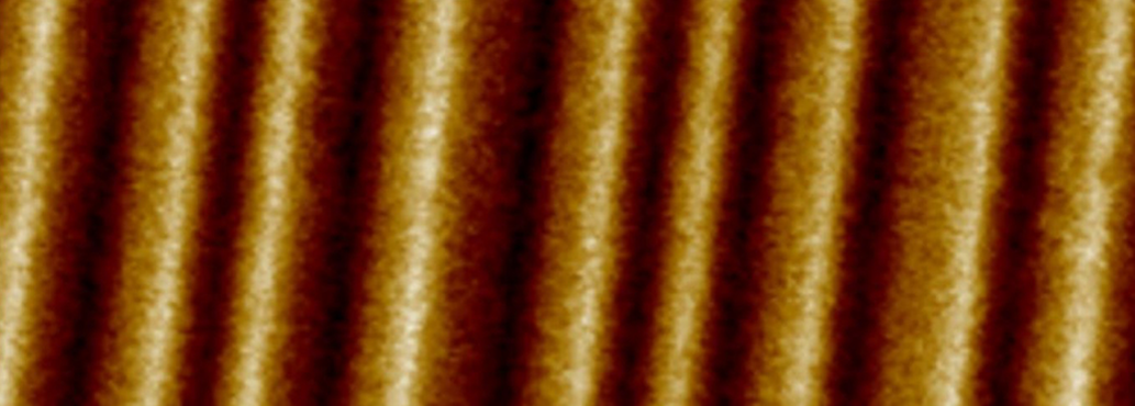
- Micro to Nanoscale Imaging and Measuring
- Nanoscale Quantification
- Electrical forces, magnetic forces
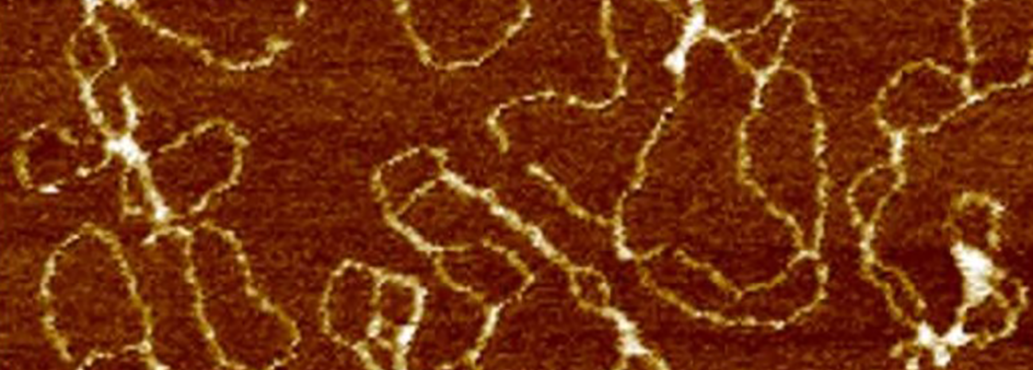
- Mechanical properties of cells, tissues, microorganisms, viruses, biological macromolecules
- Molecular biology, cell biology, and medicine
- Molecular engineering
- Solid-state physics, semiconductor science and technology
- Polymer chemistry and physics, surface chemistry
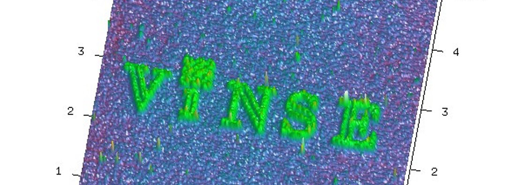
- Nanolithography
- Nanomanipulation
Contact
-

Dr. Dmitry Koktysh
VINSE Advanced Imaging
- 023 Engineering Science Building