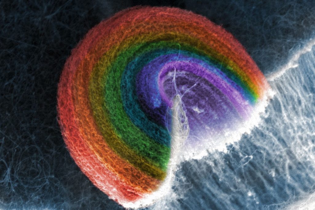
Advanced Imaging Suite

Our Advanced Imaging Suite hosts advanced electron and atomic force microscopes in spaces engineered to minimize ambient noise, vibration and, electromagnetic field levels for the best possible imaging conditions.

VINSE staff are on-site to help you get the most out of the microscopes, with easy-to-follow procedures and constant effort to develop or improve each tool’s advanced capabilities.

placeholder image
Section Contents

Equipment

User Fees

  • Bruker AFM
    • Vanderbilt personnel hourly rate: $40
    • VUMC, Fisk, TSU and Meharry hourly rate: $40
    • External universities & non-profit partners hourly rate: $44
    • Industry and external for-profit partners hourly rate: $65
  • Helios FIB/SEM
    • Vanderbilt personnel hourly rate: $70
    • VUMC, Fisk, TSU and Meharry hourly rate: $70
    • External universities & non-profit partners hourly rate: $77
    • Industry and external for-profit partners hourly rate: $179
  • Merlin SEM
    • Vanderbilt personnel hourly rate: $55
    • VUMC, Fisk, TSU and Meharry hourly rate: $55
    • External universities & non-profit partners hourly rate: $60
    • Industry and external for-profit partners hourly rate: $90
  • Osiris S/TEM
    • Vanderbilt personnel hourly rate: $70
    • VUMC, Fisk, TSU and Meharry hourly rate: $70
    • External universities & non-profit partners hourly rate: $77
    • Industry and external for-profit partners hourly rate: $192
  • Professional Services

    VINSE offers staff support for users who wish to use the facilities remotely. VINSE professional staff are able to perform your measurements, imaging, and analysis in your absence for a service fee. 

    Professional Staff Hourly Service Fees:

    • Internal: $50
    • External universities & non-profit partners hourly rate: $55
    • Industry and external for-profit partners hourly rate: $80
placeholder image
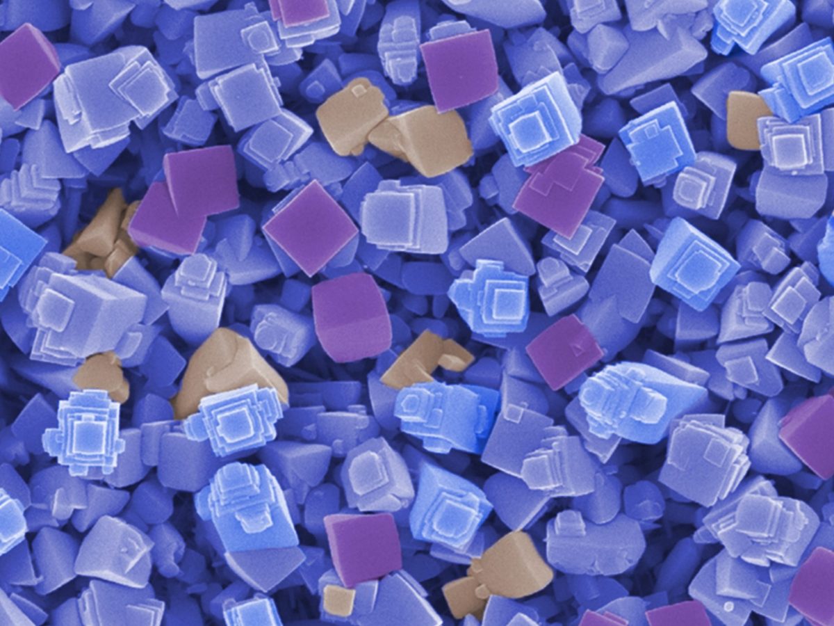
Polymethyl methacrylate (PMMA) residue on graphene after transferring to sulfonated poly-ether ether ketone (SPEEK) and soaking in acetone. Imaged in the VINSE facilities using the Zeiss Merlin Scanning Electron Microscope. (Nicole Moehring, Kidambi group)
placeholder image
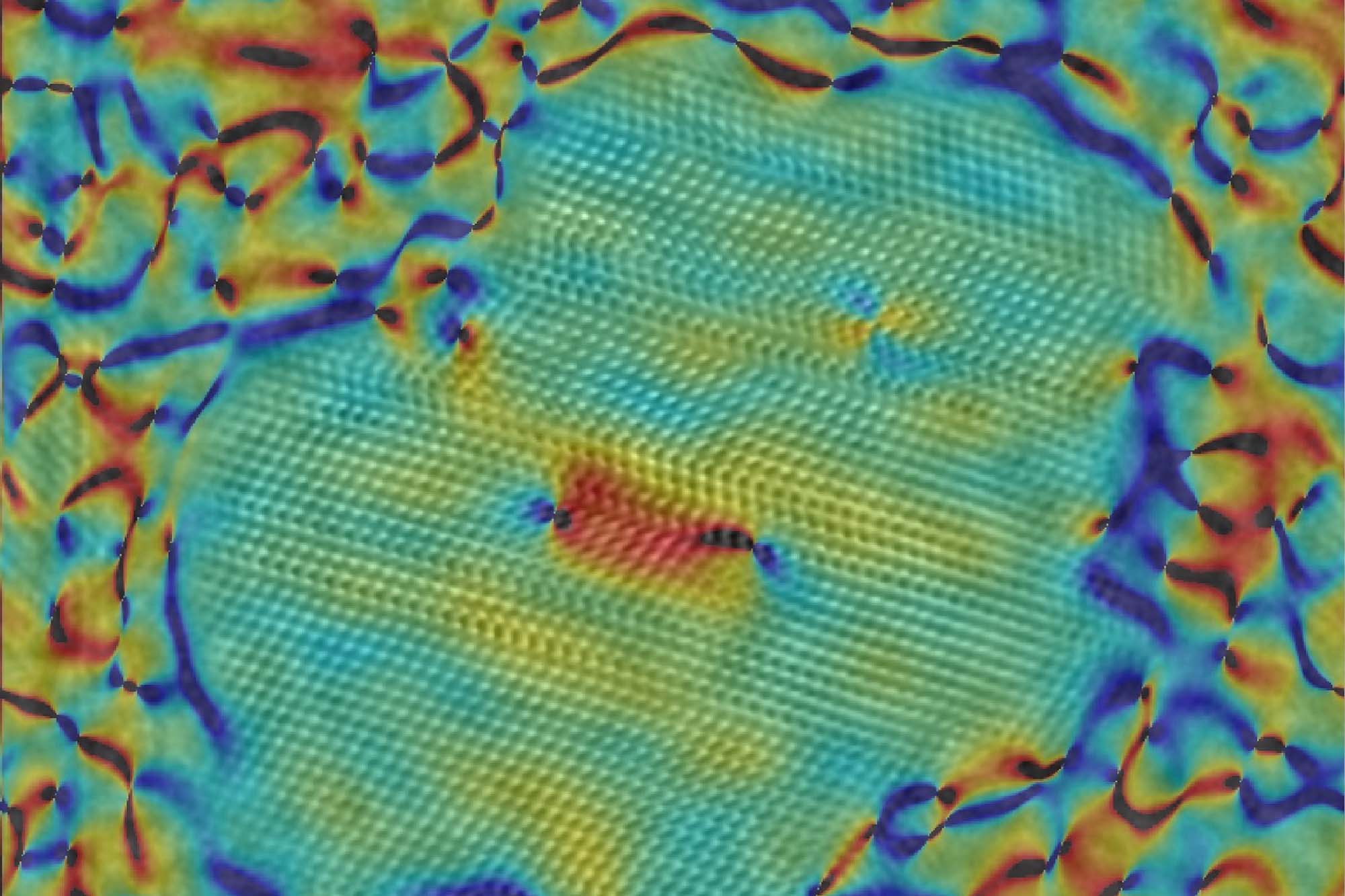
Enhanced lattice strain localized at defects in a core/shell quantum dot. Imaged in the VINSE facilities using the Tecnai Osiris Transmission Electron Microscope. (James McBride, VINSE)

Facility Details

Image
  • Millimeter to Angstrom Scale Imaging

  • Specimen Prep and Packaging Lab

  • All Sample Types

  • Cryo Capabilities

  • Nanoscale Elemental Analysis

  • User Friendly

Founded on bedrock, four imaging bays host our modern electron, ion and scanning probe instrumentation for the characterization of nanomaterials and devices.

Each bay is designed to provide a controlled environment that minimizes disturbances such as ambient noise, floor vibrations and electromagnetic field levels that meet or exceed manufacturer’s specifications for our instruments, providing the best possible imaging resolution. 
placeholder image
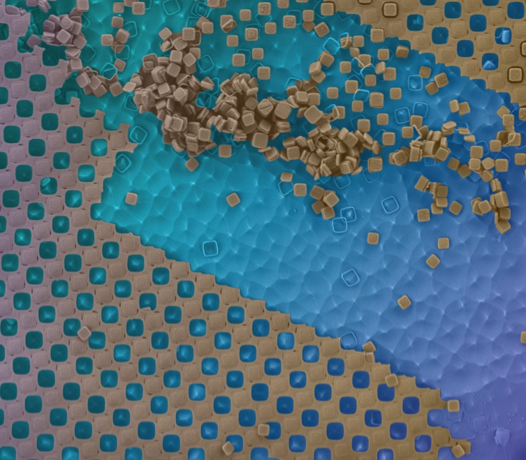
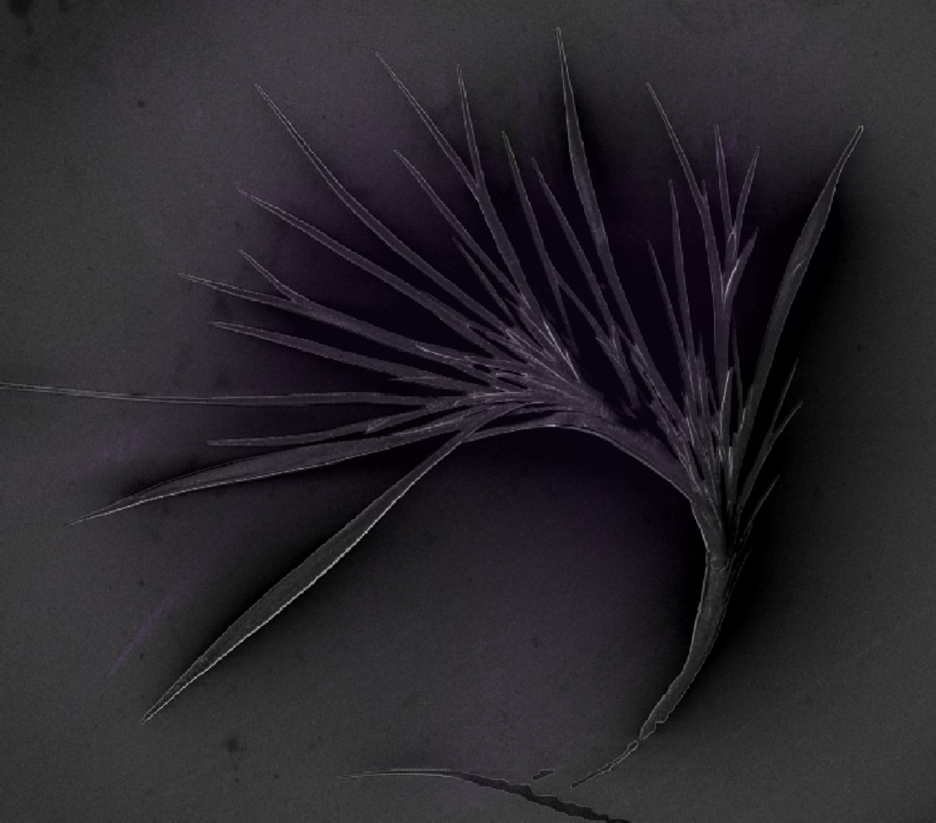
4,4′-Bis(N-carbazolyl)-1,1′-biphenyl (CBP) nanostructures on a silicon chip. Imaged in the VINSE facilities using the Zeiss Merlin Scanning Electron Microscope. (Courtney Ragle, Caldwell/Buchanan groups)
placeholder image
Photonic chip mechanically polished at Vanderbilt Earth and Environmental Science and etched at VINSE using buffered hydroflouric acid. Imaged in the VINSE facilities using the Merlin Zeiss Scanning Electron Microscope. (Kellen Arnold, Weiss group)