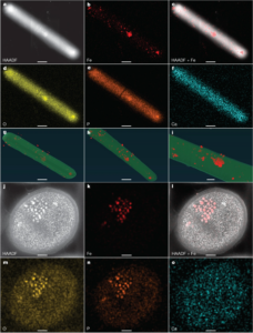
Congratulations to Eric Skaar’s group for their Nature publication. The keys towards understanding the role of nutrients in guest-hosts interactions inherently reside at the nanoscale. The work by Pi et al. from the Skaar laboratory utilized the advanced electron microscopy capabilities in VINSE to reveal how C. difficile bacteria use iron storage organelles to manage large changes in nutrient iron levels. Specifically, room temperature electron tomography in conjunction with STEM-EDS chemical imaging was performed on VINSE’s Tecnai Osiris, which enabled the nanoscale visualization and chemical identification of the bacteria-produced iron granules. Then the Helios G3 Cryo FIB-SEM in VINSE was utilized to mill thin sections of vitrified bacteria for Cryo-ET. These thin sections, prepared by Rong Sun from the Qiangjun Zhou laboratory and imaged on the Center for Structural Biology’s Titan G4 Krios, revealed that C. difficile produces membrane-bound ferrosome organelles.