New drug delivery systems, solar cells, industrial catalysts and video displays are among the potential
New drug delivery systems, solar cells, industrial catalysts and video displays are among the potential applications of special particles that possess two chemically distinct sides. These particles are named after the two-faced Roman god Janus and their twin chemical faces allow them to form novel structures and new materials.
However, as scientists have reduced the size of Janus particles down to a few nanometers in diameter – about the size of individual proteins, which has the greatest potential for drug therapy – their efforts have been hampered because they haven’t had a way to accurately map the surfaces of the particles that they produce. This uncertainty has made it difficult to evaluate the effectiveness of these particles for various applications and to improve the methods researchers are using to produce them.
Now, a team of Vanderbilt chemists has overcome this obstacle by developing the first method that can rapidly and accurately map the chemical properties of the smallest of these Janus nanoparticles.

The results, published online this month in the German chemistry journal Angewandte Chemie, address a major obstacle that has slowed the development and application of the smallest Janus nanoparticles.
The fact that Janus particles have two chemically distinct faces makes them potentially more valuable than chemically uniform particles. For example, one face can hold onto drug molecules while the other is coated with linker molecules that bind to the target cells. This advantage is greater when the different surfaces are cleanly separated into hemispheres than when the two types of surfaces are intermixed.
For larger nanoparticles (with sizes above 10 nanometers), researchers can use existing methods, such as scanning electron microscopy, to map their surface composition. This has helped researchers improve their manufacturing methods so they can produce cleanly segregated Janus particles. However, conventional methods do not work at sizes below 10 nanometers.
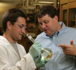
The Vanderbilt chemists – Associate Professor David Cliffel, Assistant Professor John McLean, graduate student Kellen Harkness and Lecturer Andrzej Balinski – took advantage of the capabilities of a state-of-the-art instrument called an ion mobility–mass spectrometer (IM-MS) that can simultaneously identify thousands of individual particles.

The team coated the surfaces of gold nanoparticles ranging in size from two to four nanometers with two different chemical compounds. Then they broke the nanoparticles down into clusters of four gold atoms and ran these fragments through the IM-MS.
Molecules from the two coatings were still attached to the clusters. So, by analyzing the resulting pattern, the chemists showed that they could distinguish between original nanoparticles where the two surface compounds were completely separated, those where they were randomly mixed and those that had an intermediate degree of separation.
“There is no other way to analyze structure at this scale except X-ray crystallography,” said Cliffel, “and X-ray crystallography is extremely difficult and can take months to get a single structure.”
“IM-MS isn’t quite as precise as X-ray crystallography but it is extremely practical,” added McLean, who has helped pioneer the new instrument’s development. “It can provide structural information in a few seconds. Two years ago a commercial version became available so people who want to use it no longer have to build one for themselves.”
The research was funded in part by a grant from the National Institutes of Health