Tian, Qu; Greig, Erin E.; Walker, Keenan A.; Duggan, Michael R.; Yang, Zhijian; Moghekar, Abhay; Landman, Bennett A.; Davatzikos, Christos; Resnick, Susan M.; Ferrucci, Luigi. “Longitudinal patterns of brain aging and neurodegeneration among older adults with dual decline in memory and gait.” Alzheimer’s & dementia : the journal of the Alzheimer’s Association, vol. 21, no. 2, 2025, e14612, https://doi.org/10.1002/alz.14612.
Cognitive and mobility decline together (dual decline) is more strongly linked to the risk of developing dementia than cognitive decline alone. However, it’s still unclear whether this condition is related to certain patterns of brain shrinkage, damage to white matter (the tissue that connects brain regions), or other brain changes.
In the Baltimore Longitudinal Study of Aging, we studied participants with and without dual decline to compare changes in brain images, white matter damage, and specific biomarkers over up to 13 years. We looked at brain atrophy, white matter issues, and proteins in the blood that are linked to brain health, including markers for neurodegeneration like GFAP, NfL, amyloid beta, and tau.
Our results showed that people with dual decline experienced faster shrinkage in brain regions related to memory, movement, and language. Those with only mobility problems had brain shrinkage in one specific area, while those with memory decline had a different pattern. Dual decline also showed damage to white matter in several brain areas and a greater decline in the amyloid beta ratio, which is a sign of Alzheimer’s disease. Additionally, there were higher levels of proteins linked to brain damage in the blood.
In conclusion, individuals with dual decline may be at greater risk for brain shrinkage, white matter damage, and specific changes in biomarkers that could lead to dementia. This highlights the importance of dual decline as a potential signal of underlying brain and blood changes associated with dementia.
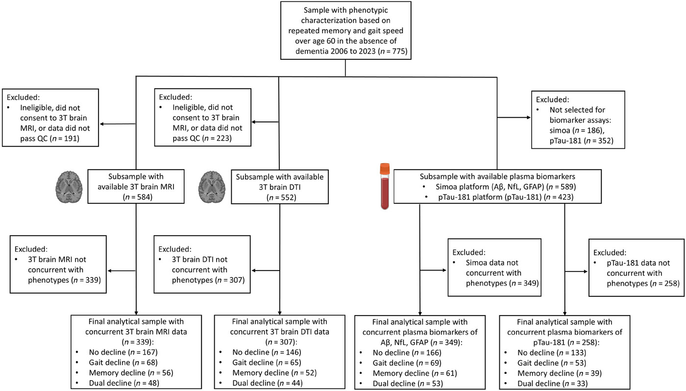
FIGURE 1
Flow chart of sample selection criteria. AD, Alzheimer’s disease; Aβ42/40, amyloid beta 42/40 ratio; DTI, diffusion tensor imaging; GFAP, glial fibrillary acidic protein; MRI, magnetic resonance imaging; NfL, neurofilament light chain; pTau181, phosphorylated tau.
