Schilling, Kurt G.; Howard, Amy F. D.; Grussu, Francesco; Ianus, Andrada; Hansen, Brian; Barrett, Rachel L. C.; Aggarwal, Manisha; Michielse, Stijn; Nasrallah, Fatima; Syeda, Warda; Wang, Nian; Veraart, Jelle; Roebroeck, Alard; Bagdasarian, Andrew F.; Eichner, Cornelius; Sepehrband, Farshid; Zimmermann, Jan; Soustelle, Lucas; Bowman, Christien; Tendler, Benjamin C.; Hertanu, Andreea; Jeurissen, Ben; Verhoye, Marleen; Frydman, Lucio; van de Looij, Yohan; Hike, David; Dunn, Jeff F.; Miller, Karla; Landman, Bennett A.; Shemesh, Noam; Anderson, Adam; McKinnon, Emilie; Farquharson, Shawna; Dell’Acqua, Flavio; Pierpaoli, Carlo; Drobnjak, Ivana; Leemans, Alexander; Harkins, Kevin D.; Descoteaux, Maxime; Xu, Duan; Huang, Hao; Santin, Mathieu D.; Grant, Samuel C.; Obenaus, Andre; Kim, Gene S.; Wu, Dan; Le Bihan, Denis; Blackband, Stephen J.; Ciobanu, Luisa; Fieremans, Els; Bai, Ruiliang; Leergaard, Trygve B.; Zhang, Jiangyang; Dyrby, Tim B.; Johnson, G. Allan; Cohen-Adad, Julien; Budde, Matthew D.; Jelescu, Ileana O. “Considerations and recommendations from the ISMRM Diffusion Study Group for preclinical diffusion MRI: Part 3—Ex vivo imaging: Data processing, comparisons with microscopy, and tractography.” Magnetic Resonance in Medicine, 2025, https://doi.org/10.1002/mrm.30424.
Preclinical diffusion MRI (dMRI) is a valuable tool for studying tissue structure, brain connectivity, and the biological processes behind diffusion. While dMRI is commonly used for non-invasive in vivo imaging (scanning living subjects), ex vivo dMRI—where tissue samples are studied outside the body—has become increasingly useful for examining tissue at a very detailed level. Ex vivo dMRI offers advantages like high-resolution images, better signal quality, and the ability to directly compare with tissue samples using histology, a method for examining tissue structure under a microscope. However, there are many challenges to consider when conducting ex vivo dMRI experiments. The process involves several complex steps, including tissue preparation, image acquisition, data processing, and interpreting results. These steps differ significantly from in vivo imaging and can influence the kinds of questions that can be answered with the data. This paper is the third part of a series that provides recommendations for preclinical dMRI studies. It outlines best practices for preparing and processing ex vivo tissue images, with a focus on data handling and comparison with microscopic analysis. We offer guidelines where possible, discuss areas that still lack clear guidelines, and suggest future directions for research. Finally, we encourage the sharing of code and data and highlight open-source software and databases that are specifically useful for small animal and ex vivo imaging.
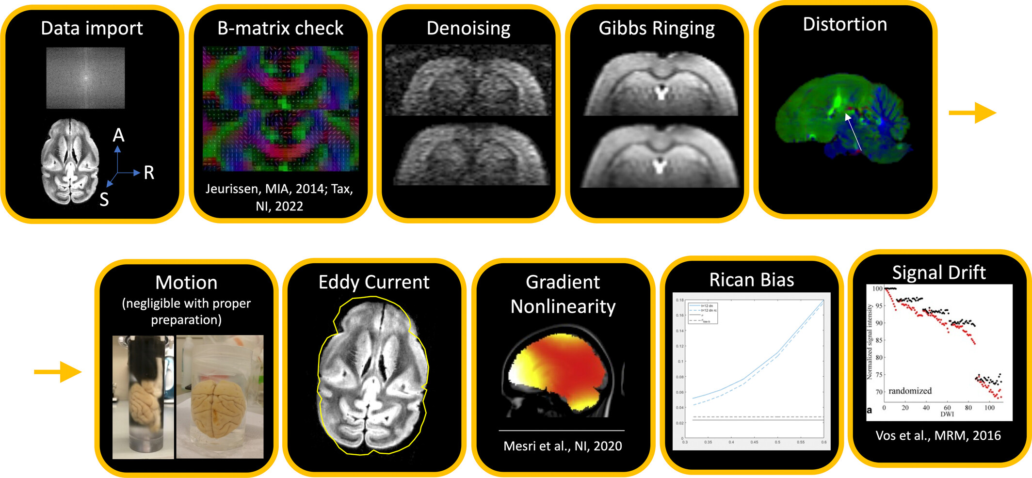
FIGURE 1
There are many artifacts that must be corrected for in preprocessing. These are not necessarily presented in order, and correction may not be necessary in all cases. Nevertheless, the most common order for pre-processing steps (after data important and quality check) is: (i) thermal noise reduction (referred to as denoising), (ii) Gibbs ringing correction, (iii) susceptibility distortion + motion + eddy current corrections (+ gradient non-linearity, if applicable), (iv) Rician bias correction and (v) signal drift correction. Figures kindly provided by Ileana Jelescu, Kurt Schilling, or reproduced from.2, 3