Rasdall, Marselle A.; Cho, Chloe; Stahl, Amy N.; Tovar, David A.; Lavin, Patrick; Kerley, Cailey I.; Chen, Qingxia; Ji, Xiangyu; Colyer, Marcus H.; Groves, Lucas; Longmuir, Reid; Chomsky, Amy; Gallagher, Martin J.; Anderson, Adam; Landman, Bennett A.; Rex, Tonia S. “Primary Visual Pathway Changes in Individuals With Chronic Mild Traumatic Brain Injury.” JAMA ophthalmology, vol. 143, no. 1, 2025, pp. 33-42, https://doi.org/10.1001/jamaophthalmol.2024.5076.
Many people with mild traumatic brain injury (TBI) experience vision problems, even when standard eye exams show no issues. However, there is no widely accepted method for diagnosing these subtle visual impairments. This study explored whether a combination of vision tests and machine-learning techniques could help detect visual dysfunction in people with mild TBI.
Between 2018 and 2021, researchers conducted a study at a trauma research hospital, comparing 28 individuals with a history of mild TBI to 28 people with no history of brain injury. All participants had normal vision according to standard eye tests. The study excluded individuals with moderate or severe TBI, eye diseases, neurological or psychiatric conditions, or other factors that could affect vision. Participants underwent a series of assessments, including eye movement tests, contrast sensitivity measurements, brain scans, and visual field testing.
The results revealed subtle but significant differences in the visual systems of those with mild TBI. They had difficulty coordinating their eyes, reduced ability to see contrasts between objects, and changes in how their brains processed visual information. Some participants also showed differences in the structure of their optic nerves and brain regions involved in vision. Using machine-learning techniques, researchers detected additional brain changes related to vision, even in individuals who did not report any vision problems.
These findings suggest that mild TBI can affect vision in ways that traditional eye exams may not detect. A combination of specialized tests and advanced technology, such as machine learning, could help improve the diagnosis of visual problems in individuals with mild TBI, leading to better treatment and care.
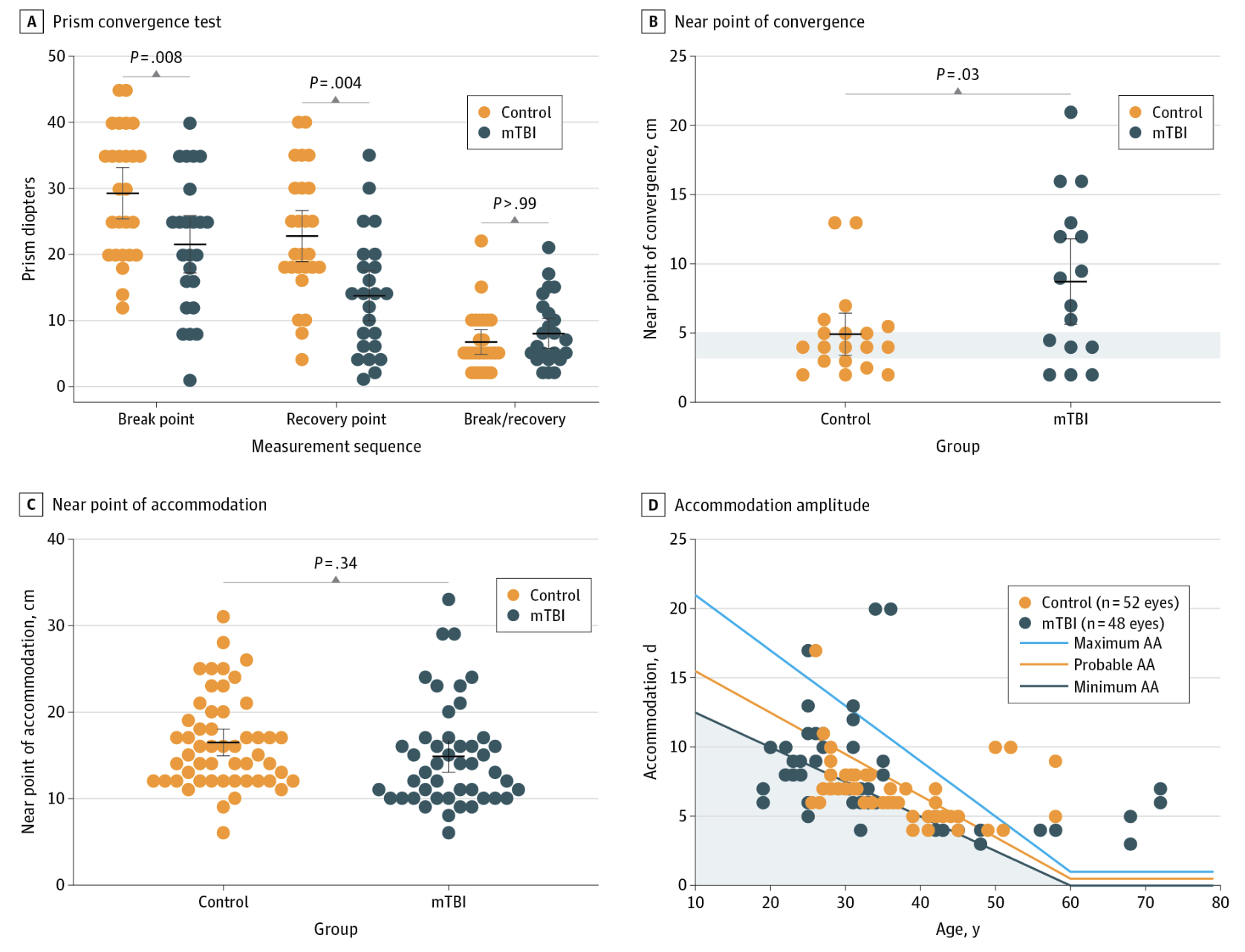
Figure 1 : A, Quantification of prism convergence test (PCT), represented as diopters for break point, recovery point, and prism fusion range. B, Quantification of the near point of convergence (NPC), with the normal range shown in the shaded gray area derived from age- and sex-adjusted normative data; values greater than 6 cm are considered abnormal. C, Quantification of near point of accommodation (NPA). D, NPA values were converted to diopters and plotted against age, overlaid with the Hofstetter equation. Horizontal lines between the upper and lower whisker marks indicate the mean, and the top and bottom whisker marks represent the 95% CIs. A 2-sided P value threshold of .01 was set for PCT and NPC, and .03 for NPA, using a Bonferroni correction to control for multiple comparisons, with a type I error rate of 0.05 per measurement. Analyses were conducted using a linear mixed-effects model and the Wilcoxon rank sum test. AA indicates accommodation amplitude; mTBI, mild traumatic brain injury.
