Lee, Ho Hin; Saunders, Adam M.; Kim, Michael E.; Remedios, Samuel W.; Remedios, Lucas W.; Tang, Yucheng; Yang, Qi; Yu, Xin; Bao, Shunxing; Cho, Chloe; Mawn, Louise A.; Rex, Tonia S.; Schey, Kevin L.; Dewey, Blake E.; Spraggins, Jeffrey M.; Prince, Jerry L.; Huo, Yuankai; Landman, Bennett A. “Super-resolution multi-contrast unbiased eye atlases with deep probabilistic refinement.” Journal of Medical Imaging, vol. 11, no. 6, 2024, 64004, https://doi.org/10.1117/1.JMI.11.6.064004.
The shape and structure of eyes can vary significantly from person to person, especially in areas like the eye socket and optic nerve. These variations make it difficult to create a standard, accurate map of the eye that works for everyone. To address this challenge, we propose a new method for creating detailed and unbiased eye maps. First, we use a deep learning technique to improve the quality of scans that lack detail, turning lower-resolution images into clearer ones. We then create an initial reference map using scans from a small group of people and adjust additional scans from other individuals to fit this reference map. To refine the map further, we apply an advanced machine learning technique, which helps better align the boundaries of eye structures. This approach was tested using MRI scans, creating four different eye maps based on various tissue types.
The results showed that by refining the reference map with enough scans, our method significantly improved the accuracy of how the eye’s structures were aligned, compared to traditional methods. This improvement was confirmed through statistical tests, demonstrating that our approach better matched the shapes of eye organs. In conclusion, by combining techniques like improving scan quality and using deep learning models, we have developed a more accurate and standardized way to create eye maps that can be applied to a diverse population. This method overcomes the natural variation in eye shapes and provides a reliable reference for both research and medical applications.
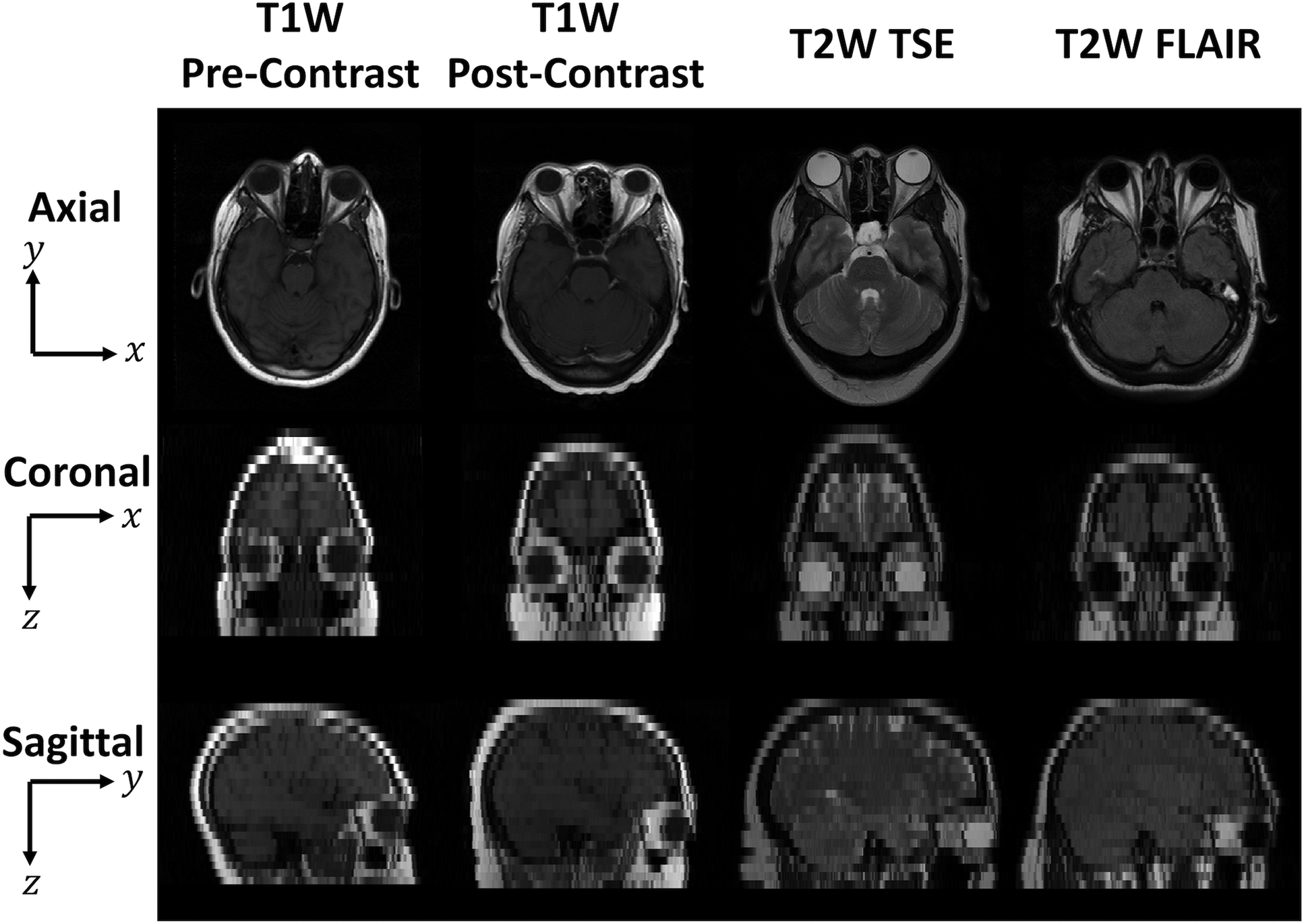
Fig. 1
Representative in-plane (axial, first row) and through-plane (coronal, second row and sagittal, third row) slices for four MRI tissue contrast from four different subjects. The coronal and sagittal through-plane slices are lower resolution than the axial in-plane slices and are visualized with nearest-neighbor interpolation. The relatively lower resolution limits our ability to distinguish organs and generalize anatomical characteristics across populations.
