
Tian, Qu; Greig, Erin E.; Davatzikos, Christos; Landman, Bennett A.; Resnick, Susan M.; Ferrucci, Luigi. “Higher skeletal muscle mitochondrial oxidative capacity is associated with preserved brain structure up to over a decade.” Nature Communications, vol. 15, no. 1, 2024, 10786, https://doi.org/10.1038/s41467-024-55009-z.
Impaired muscle function, specifically related to the mitochondria in muscles, has been linked to future cognitive decline and higher levels of Alzheimer’s disease and neurodegeneration biomarkers. This study looks at how muscle oxidative capacity (the ability of muscles to use oxygen efficiently) is associated with changes in brain structure over more than a decade. We found that higher muscle oxidative capacity, measured by MR spectroscopy after exercise recovery, is linked to less enlargement of the brain’s ventricles (fluid-filled spaces) and slower brain aging. Additionally, it was associated with less shrinkage in key brain regions, such as the sensorimotor cortex, parts of the temporal and occipital lobes, the thalamus, and areas involved in emotion and motor control. We also observed that higher muscle oxidative capacity was connected to better-preserved white matter, which is essential for communication between different brain regions, particularly in the cingulate area, corpus callosum, and superior longitudinal fasciculus. Overall, higher muscle oxidative capacity helps preserve brain structure over time, especially in areas critical for thinking, movement, and sensory processing.
Fig. 1: Flow chart of the selection of study sample.
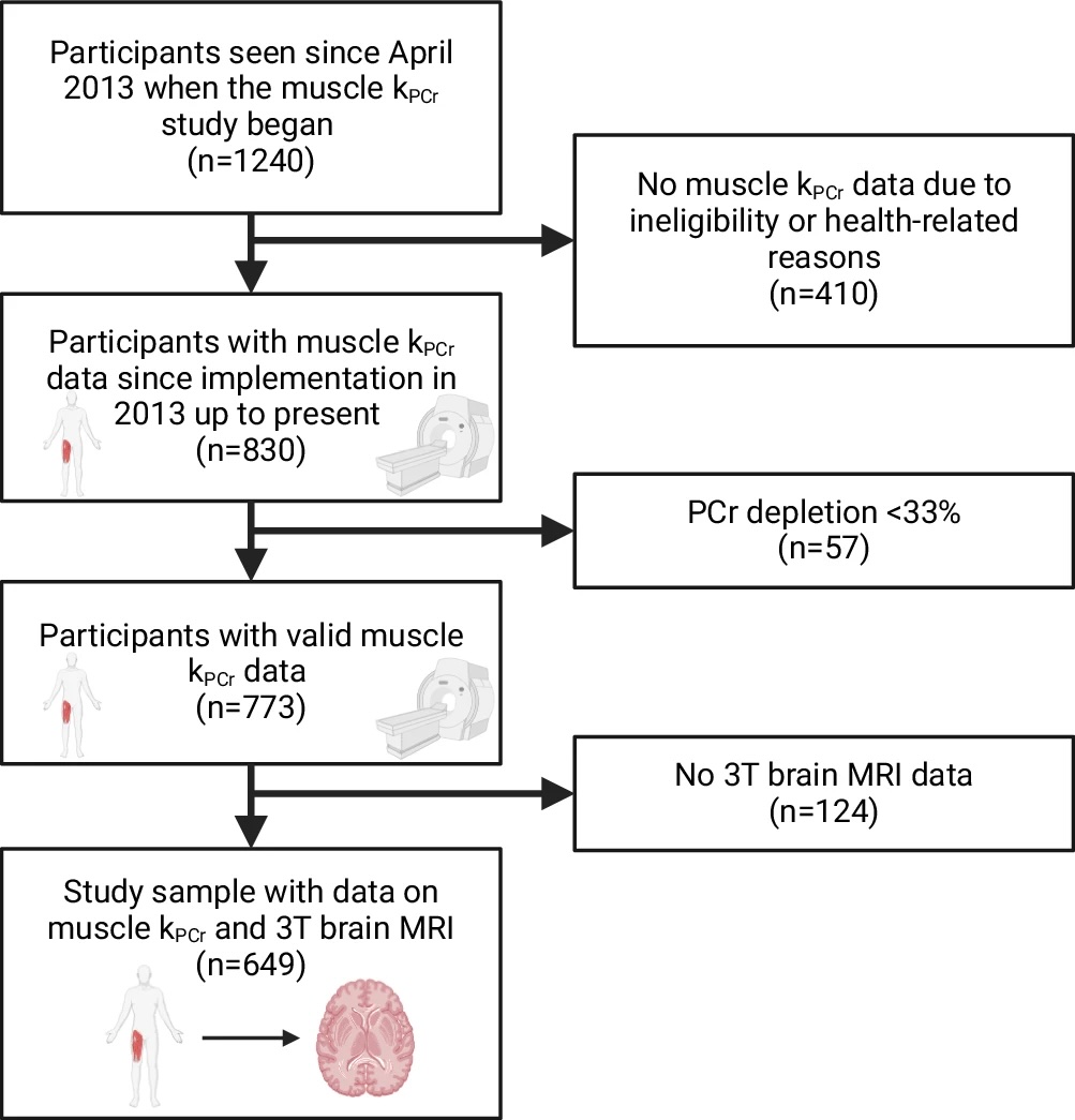
Legend: MRI magnetic resonance imaging. Created in BioRender. Greig, E. (2024) https://BioRender.com/k39x234. kPCr stands for the post-exercise recovery rate of phosphocreatine (PCr).