Trujillo, Paula; O’Rourke, Kaitlyn R.; Roman, Olivia C.; Song, Alexander K.; Hett, Kilian; Cooper, Amy; Black, Bonnie K.; Donahue, Manus J.; Shibao, Cyndya A.; Biaggioni, Italo; Claassen, Daniel O. “Central Involvement in Pure Autonomic Failure: Insights from Neuromelanin-Sensitive Magnetic Resonance Imaging and 18F-Fluorodopa-Positron Emission Tomography.” Movement Disorders, 2025, https://doi.org/10.1002/mds.30119.
Central synucleinopathies, which include conditions like Parkinson’s disease (PD), dementia with Lewy bodies (DLB), and multiple system atrophy (MSA), are characterized by a buildup of a protein called alpha-synuclein and loss of certain brain cells. These conditions often begin with symptoms in the brain’s substantia nigra (SN) and locus coeruleus (LC). Another condition, known as pure autonomic failure (PAF), affects the nervous system outside the brain and can sometimes be an early warning sign of these central synucleinopathies.
The goal of this study was to explore early changes in the brain in patients with PAF using advanced imaging techniques, specifically neuromelanin-sensitive magnetic resonance imaging (NM-MRI) and fluorodopa-positron emission tomography (FDOPA-PET). The study aimed to find out whether people with PAF who are likely to develop central synucleinopathies show changes in the contrast in the LC and SN regions of the brain compared to healthy individuals or those less likely to progress to these conditions.
The study included 23 PAF patients, divided into two groups: those at high risk (13) and low risk (10) of developing a central synucleinopathy. The study also included 22 PD patients, 8 DLB patients, and 23 healthy controls for comparison. Using NM-MRI and FDOPA-PET scans, the researchers measured brain activity and dopamine production in different regions.
The results showed that the high-risk PAF patients had less contrast in the LC and SN regions of the brain compared to healthy controls and low-risk PAF patients. Their results were similar to those observed in PD and DLB patients. In fact, the level of contrast in the SN was related to how much dopamine the brain was using. The study also included follow-up scans of 6 PAF patients, which showed that those who developed central synucleinopathies had reduced NM-MRI and PET values over time.
In conclusion, both NM-MRI and FDOPA-PET could be useful tools for detecting early changes in the brain and predicting the progression of PAF to more severe synucleinopathies. These imaging techniques could help doctors identify patients who may benefit from early intervention.
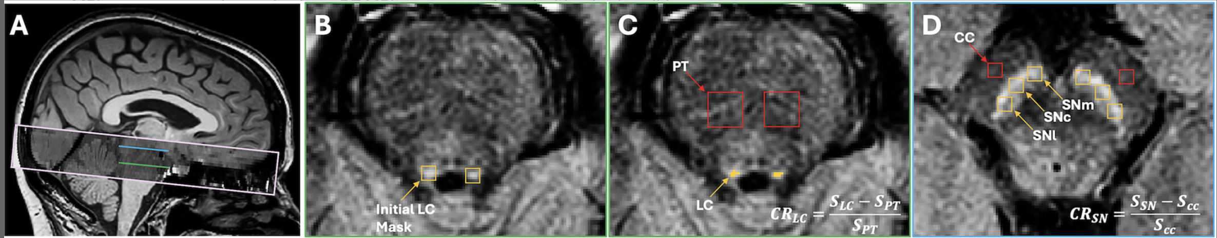
FIG. 1
Definition of the regions of interest (ROIs) for the locus coeruleus (LC) and substantia nigra (SN). (A) Sagittal slice of the anatomical T1-weighted image showing the field-of-view coverage for the neuromelanin-sensitive magnetic resonance imaging (NM-MRI), oriented orthogonally to the floor of the fourth ventricle and spanning between the superior colliculi and the superior cerebellar peduncles (pink rectangle). The corresponding axial views for the LC (green line) and SN (blue line) are depicted in B–D. (B) Initial masks around the LC. (C) Final LC ROI, consisting of the seven contiguous voxels (per slice and side) with the highest signal intensity within the initial mask, and a reference ROI in the pontine tegmentum (PT). (D) Reference region in the cerebral crus (CC) and the lateral (SNl), central (SNc), and medial (SNm) subregions of the SN. [Color figure can be viewed at wileyonlinelibrary.com]