Xu, L.; Zhao, Y.; Choi, S.; Li, M.; Schilling, K.G.; Zu, Z.; Rogers, B.P.; Ding, Z.; Anderson, A.W.; Landman, B.A.; Gore, J.C.; Gao, Y. “Reductions in the white–gray functional connectome in preclinical Alzheimer’s disease and their associations with amyloid and cognition.” Alzheimer’s and Dementia, 2024, DOI: 10.1002/alz.14334.
This study explored how the brain’s connections between white and gray matter (WM-GM) are altered in early (preclinical) Alzheimer’s disease (AD) and how these changes relate to amyloid buildup and thinking abilities. Researchers analyzed brain connectivity and network properties in 344 participants, including those with preclinical AD, AD dementia, and healthy controls.
They found that preclinical AD is linked to weaker connections in specific WM-GM regions and less separation of brain networks related to control, attention, and movement. Many of these changes were tied to higher levels of amyloid beta, a hallmark of AD. One specific connection showed a direct link to cognitive decline. In AD dementia, these disruptions were more widespread and strongly associated with amyloid levels and cognitive issues. These findings highlight how changes in WM-GM connectivity offer insights into brain dysfunction early in the AD process.

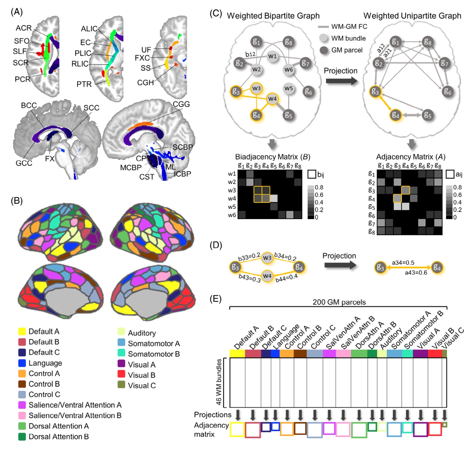
FIGURE 1
Atlas-defined WM bundles and GM parcels, illustration of bipartite graph model, split of biadjacency matrix into functional
networks. A, Forty-six atlas-defined WM bundles (Table S2 in supporting information). B, Two hundred atlas-defined GM parcels are classified into 17 functional networks (Table S3 in supporting information). C, A simplified bipartite model and the projection from weighted bipartite graph to weighted unipartite graph. The biadjacency matrix is the adjacency matrix of a WM–GM FC graph. D, An example illustrating the algorithm of bipartite-to-unipartite projection. E, Split of biadjacency matrix into 17 functional networks and subsequent projections. ACR, anterior corona radiata; ALIC, anterior limb of internal capsule; BCC, body of corpus callosum; CGG, cingulum in the cingulate gyrus; CGH, cingulum hippocampus; CP, cerebral peduncle; CST, corticospinal tract; EC, external capsule; FC, functional connectivity; FX, fornix; FXC, fornix cres; GCC, genu of corpus callosum; GM, gray matter; ICBP, inferior cerebellar peduncle; MCBP, middle cerebellar peduncle; ML, medial lemniscus; PCR, posterior corona
radiata; PLIC, posterior limb of internal capsule; PTR, posterior thalamic radiation; RLIC, retrolenticular limb of internal capsule; SCBP, superior cerebellar peduncle; SCC, splenium of corpus callosum; SCR, superior corona radiata; SFO, superior fronto-occipital fasciculus; SLF, superior longitudinal fasciculus; SS, sagittal stratum; UF, uncinate fasciculus; WM, white matter