Leguizamon, M.; McKnight, C.D.; Ponzo, T.; Elenberger, J.; Eisma, J.J.; Song, A.K.; Trujillo, P.; Considine, C.M.; Donahue, M.J.; Claassen, D.O.; Hett, K. “Intravenous arachnoid granulation hypertrophy in patients with Parkinson disease.” npj Parkinson’s Disease, Volume 10, Issue 1, 2024, Article 177, DOI: 10.1038/s41531-024-00796-x.
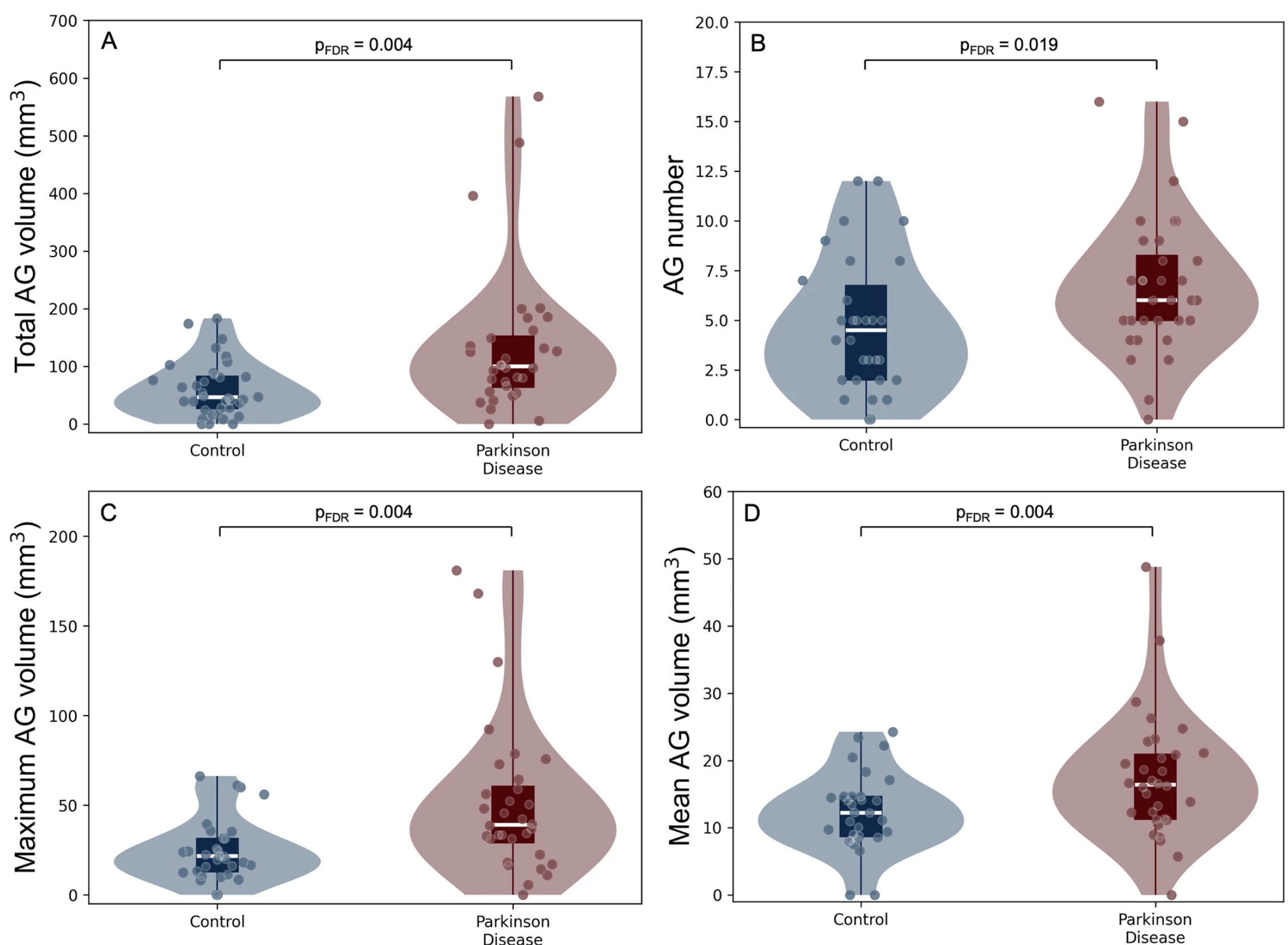
Arachnoid granulations (AGs) are small structures that help manage cerebrospinal fluid (CSF) flow. In Parkinson’s disease (PD), issues with CSF flow might relate to changes in AG size and function. This study compared AG size and number between healthy individuals and those with PD and examined their links to symptoms. Results showed that people with PD had larger and more AGs than healthy controls. These AG changes were linked to motor and cognitive symptoms and to objective sleep problems, though not to self-reported sleep issues. This suggests AGs may play a role in PD-related changes in the brain.

Fig. 1: Group differences in arachnoid granulation (AG) total volume (mm3). A Significantly increased total AG volume in baseline Parkinson disease scans compared to healthy controls. B Significantly increased AG number in Parkinson disease compared to healthy controls. CSignificantly increased maximum AG volume in Parkinson disease compared to healthy controls. DSignificantly increased mean AG volume in Parkinson disease compared to healthy controls. Violin plots are shown with conventional boxplots and individual data points overlaid. Corrected p-values are shown.