Functional correlation tensors in brain white matter and the effects of normal aging

Xu, Lyuan, Gao, Yurui, Li, Muwei, Lawless, Richard, Zhao, Yu, Schilling, Kurt G., Rogers, Baxter P., Anderson, Adam W., Ding, Zhaohua, Landman, Bennett A., & Gore, John C. (2024). Functional correlation tensors in brain white matter and the effects of normal aging. Brain Imaging and Behavior. https://doi.org/10.1007/s11682-024-00914-6

This study investigates how the brain’s white matter (WM) changes with age by examining the relationships between brain activity and WM microstructure using a method called Functional Correlation Tensors (FCTs) derived from resting-state fMRI data. FCTs are used to analyze the directionality and strength of brain activity correlations in white matter, helping researchers understand how brain communication and structure interact, especially in aging.

The researchers studied data from 461 cognitively normal adults, aged 42 to 95, sourced from a publicly available database. By looking at FCT metrics—such as how signals move along and across WM tracts—they were able to map out patterns of brain activity and measure how these change with age. The analysis showed that some areas of white matter experience declines in functional correlations (weaker communication) with age, while other areas see increases. Additionally, women appeared to show age-related changes in more brain regions compared to men, although the interaction between age and sex was not statistically significant.

This study demonstrates that FCTs follow a consistent spatial distribution across individuals, offering a reproducible way to quantify subtle changes in WM as people age. The results provide new insights into the effects of aging on brain structure and function, potentially informing future research into brain health, cognitive decline, and neurological diseases.
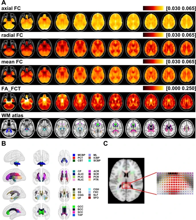
(A) Averaged adjusted maps of axial FC, radial FC, mean FC, FA_FCT across all subjects and JHU ICBM-DTI-81 WM atlas used in this work. (B) WM bundles defined by JHU ICBM-DTI-81 WM atlas. The abbreviations in the right column are: (1) Tracts in the Brainstem: MCBP: middle cerebellar peduncle; ML: medial lemniscus; PCT: pontine crossing tract; ICBP: inferior cerebellar peduncle; CST: corticospinal tract; SCBP: superior cerebellar peduncle; (2) Projection fibers: CP: cerebral peduncle; ALIC: anterior limb of internal capsule; PLIC: posterior limb of internal capsule; RLIC: retrolenticular part of the internal capsule; ACR: anterior corona radiata; SCR: superior corona radiata; PCR: posterior corona radiata; PTR: posterior thalamic radiation; (3) Association fibers: FX: fornix; SS: sagittal stratum; CGG: cingulum in the cingulate gyrus; CGH: cingulum in the hippocampus; FXC: fornix (cres); SLF: superior longitudinal fasciculus; SFO: superior fronto-occipital fasciculus; UF: uncinate fasciculus; (4) Commissural fibers: GCC: genu of corpus callosum; BCC: body of corpus callosum; SCC: splenium of corpus callosum; TAP: tapetum. (C) FCT ellipsoid map of a subject weighted by its own FCT FA and localized magnification image of the corpus callosum region.

Explore Story Topics