
Tian Yu, Yunhe Li, Michael E. Kim, Chenyu Gao, Qi Yang, Leon Y. Cai, Susan M. Resnick, Lori L. Beason-Held, Daniel C. Moyer, Kurt G. Schilling, and Bennett A. Landman. “Tractography with T1-weighted MRI and Associated Anatomical Constraints on Clinical Quality Diffusion MRI.” Proceedings of SPIE Medical Imaging 2024: Image Processing, vol. 12926, 129262B, 2024, San Diego, California
Diffusion MRI (dMRI) streamline tractography is the gold standard for estimating brain white matter (WM) pathways in vivo, traditionally reflecting macroscopic relationships with WM microstructure. However, recent advancements have shown that convolutional recurrent neural networks (CoRNN), trained with a teacher-student framework, can learn and propagate streamlines directly from T1 and anatomical contexts. Previously, training for these networks required high-resolution dMRI data.
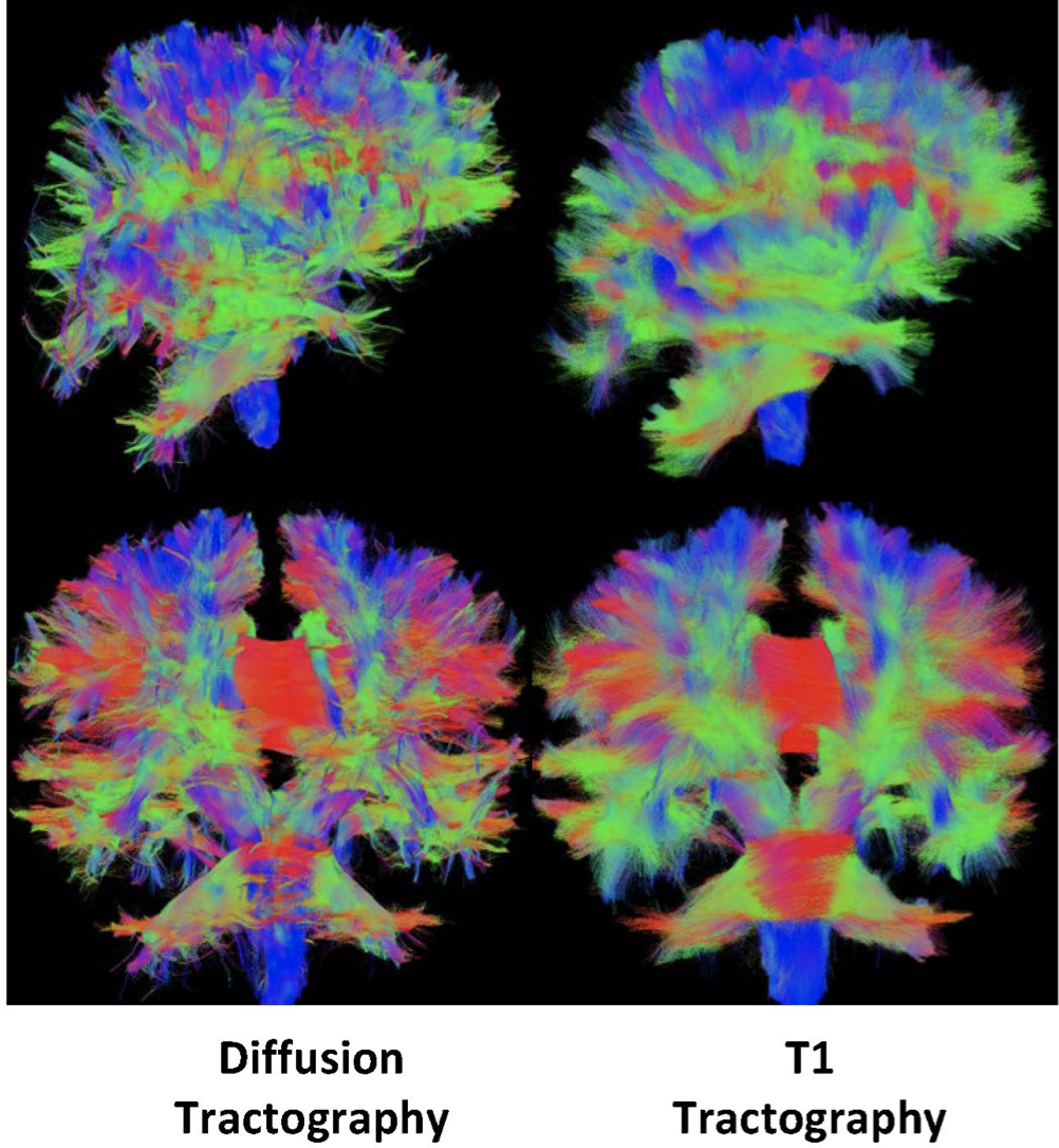
In this study, the training mechanism is generalized to work with traditional clinical resolution data, enhancing applicability across diverse and sensitive populations. CoRNN was trained on a subset of the Baltimore Longitudinal Study of Aging (BLSA), which closely resembles typical clinical protocols. A new metric, the epsilon ball seeding method, was introduced to compare T1-based tractography with traditional diffusion tractography at the streamline level. Using this metric, it was found that T1 tractography generated by CoRNN replicates diffusion tractography with an error margin of approximately two millimeters.
These findings suggest that CoRNN can effectively generalize across different data resolutions, making T1-based tractography a viable alternative to traditional diffusion tractography in clinical settings.
been only done with dMRI data. Recent work has shown that similar structures can be learned from only T1 images and
the anatomical context they provide. Visually the connections are incredibly similar. No prior work has explored the
difference on a streamline-by-streamline basis.