Nagasoujanya V. Annasamudram, Azubuike M. Okorie, Richard G. Spencer, Rita R. Kalyani, Qi Yang, Bennett A. Landman, Luigi Ferrucci, and Sokratis Makrogiannis. “Multi-method and Multi-atlas Segmentation Fusion for Delineation of Thigh Muscle Groups in 3D Water-Fat Separated MRI.” Proceedings of SPIE Medical Imaging 2024: Image Processing, vol. 12926, 129262X, 2024, San Diego, California,
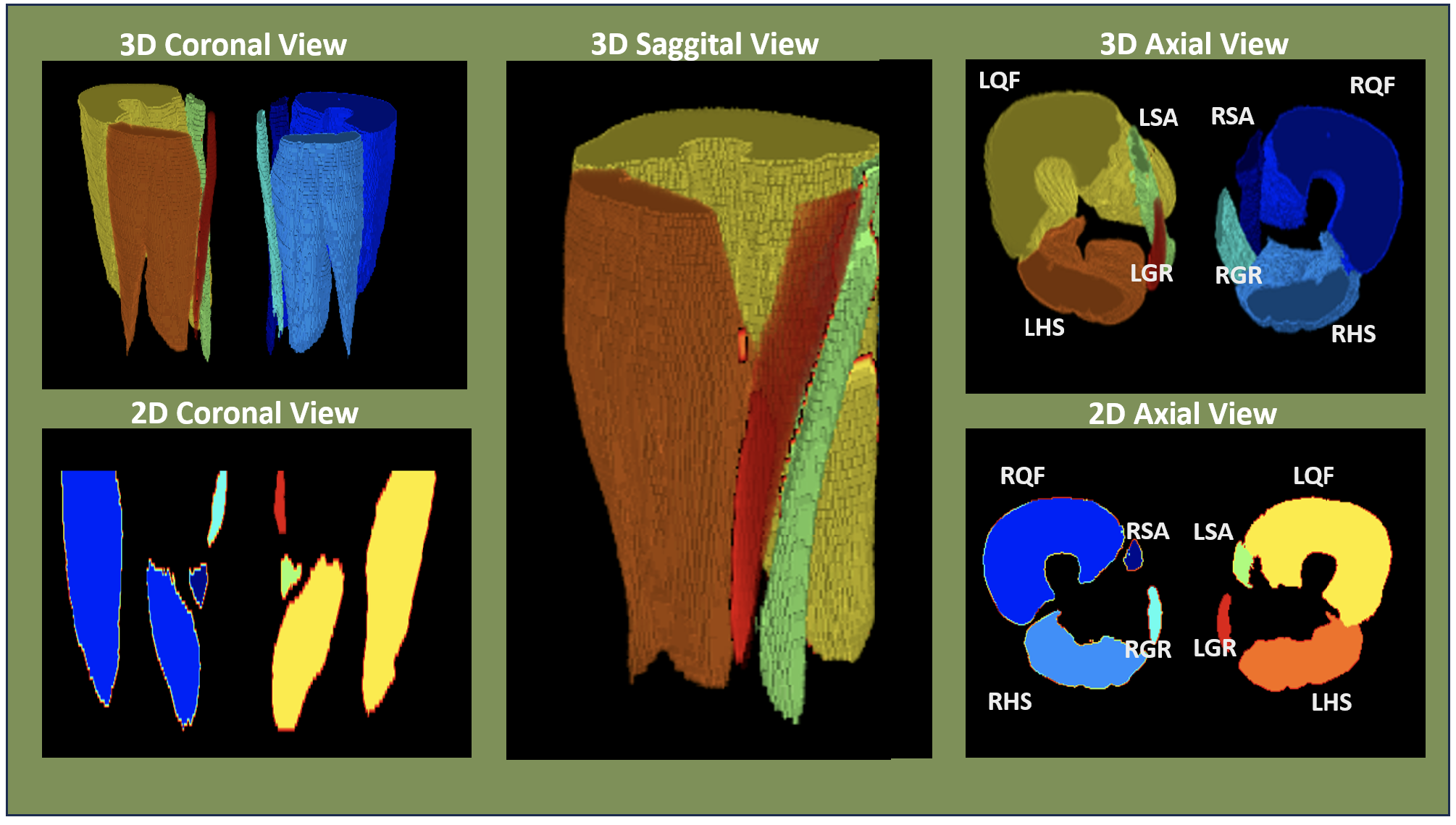
Segmentation is a crucial technique for analyzing tissue properties, with uses in body composition assessment, disease diagnosis, and the development of imaging biomarkers. This study introduces a new automated method for segmenting functional muscle groups in 3D MRI scans of the mid-thigh. Segmenting these muscle groups is challenging due to their close anatomical proximity. The proposed method combines multiple techniques and reference atlases to enhance accuracy. It uses anatomical mappings to better differentiate adjacent muscle groups, improving segmentation where conventional methods fall short.
The researchers segmented four muscle groups in both thighs using this multi-atlas approach, which merges results from different anatomical reference points. They tested four different deformable models—free-form deformation (FFD), symmetric normalization (SYN), symmetric diffeomorphic demons (SDD), and Voxelmorph (VXM)—and combined these methods for optimal results. Their approach achieved an average Dice Similarity Coefficient (DSC) of 0.795, indicating high accuracy in delineating muscle groups. This method shows promise for improving muscle segmentation in medical imaging.