
Alexa L. Eby, Lucas W. Remedios, Michael E. Kim, Muwei Li, Yurui Gao, John C. Gore, Kurt G. Schilling, and Bennett A. Landman. “Identification of Functional White Matter Networks in BOLD fMRI.” Proceedings of SPIE Medical Imaging 2024: Image Processing, vol. 12926, 129260T, 2024, San Diego, California, United States
White matter signals in resting-state blood oxygen level dependent (BOLD) functional magnetic resonance imaging (fMRI) have often been overlooked, yet increasing evidence suggests that these signals are indicative of brain activity. Understanding how these white matter signals capture function can provide valuable insights into brain physiology and potentially serve as early markers for neurological changes, such as those seen in Alzheimer’s Disease.
To explore white matter brain networks, researchers used the OASIS-3 dataset to extract white matter signals from resting-state BOLD-fMRI data from 711 subjects, resulting in a total of 2,026 images. The imaging was longitudinal, allowing for the examination of changes over time. Hierarchical clustering was applied to investigate clusters of voxel-level correlations in the timeseries data. The stability of these clusters was measured using average Dice coefficients across two different cross-validation methods: one validating stability between scans and the other between subject populations.
The study found that functional clusters at hierarchical levels 4, 9, 13, 18, and 24 exhibited local maximum stability, indicating better-defined white matter clusters. When compared with regions defined by the JHU-DTI-SS Type-I Atlas, clusters at lower hierarchical levels corresponded well with anatomical lobes. At higher hierarchical levels, functional clusters identified motor and memory regions, covering 50.00%, 20.00%, 27.27%, and 35.14% of the frontal, occipital, parietal, and temporal lobes, respectively.
These findings suggest that white matter signals in resting-state BOLD-fMRI can provide meaningful information about brain function and anatomy, highlighting their potential role in studying brain activity and neurological changes.
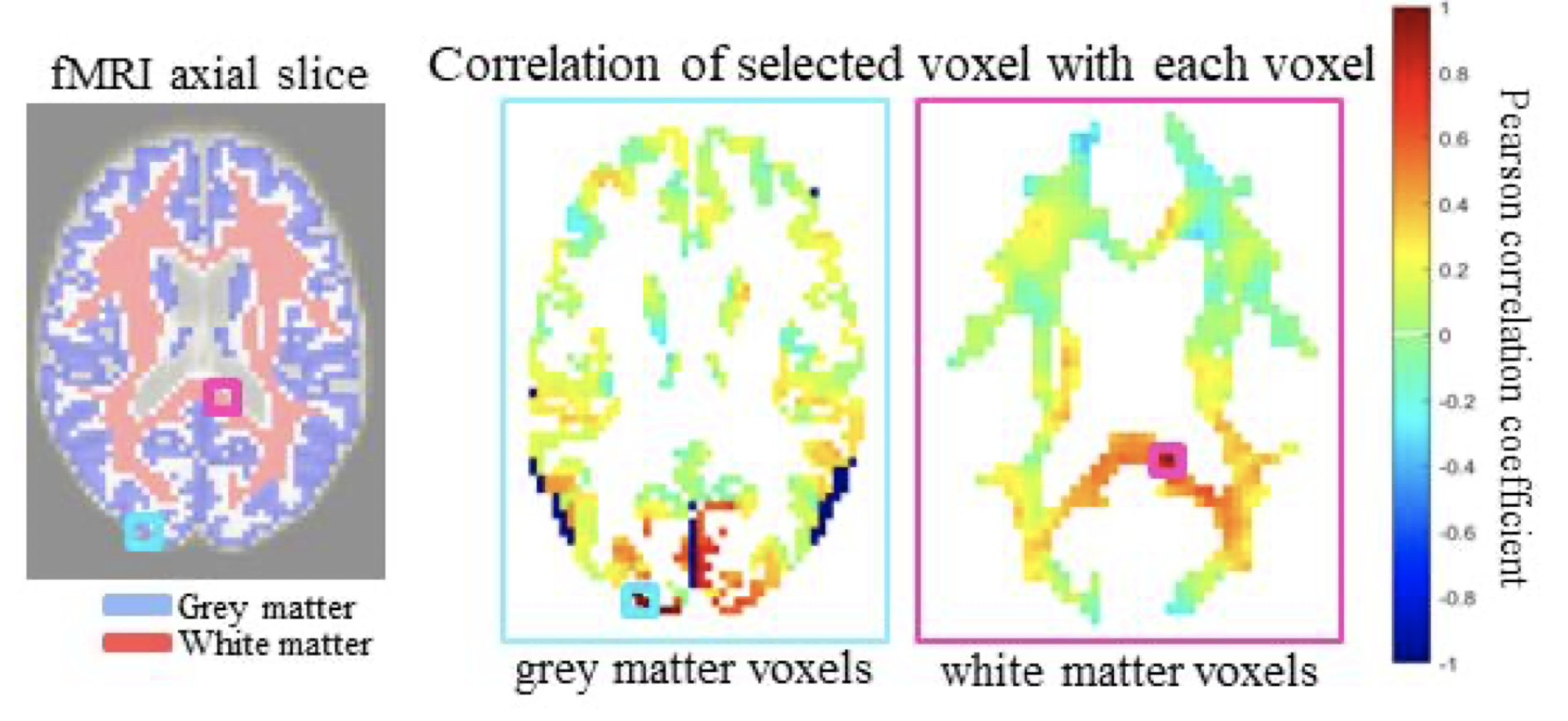
identify anatomical regions7 (left). In a similar manner, we hypothesize correlations can be identified between white matter voxels
(right), providing evidence there may be informative white matter correlations in resting state BOLD fMRI data.