
Ema Topolnjak, Chenyu Gao, Lori L. Beason-Held, Susan M. Resnick, Kurt G. Schilling, and Bennett A. Landman. “Assessment of Subject Head Motion in Diffusion MRI.” Proceedings of SPIE Medical Imaging 2024: Image Processing, vol. 12926, 129261B, 2024, San Diego, California
Subject head motion during the acquisition of diffusion-weighted imaging (DWI) of the brain can induce artifacts and negatively affect image quality. Understanding the frequency and extent of motion can highlight the most critical aspects of motion correction needed. This study investigates the extent of translational and rotational movements among participants and examines how these motions change during scan acquisition.
The analysis includes 5,380 DWI scans from 1,034 participants. The study measures rotations and translations in the sagittal, coronal, and transverse planes required to align each volume to the first and previous volumes, as well as overall displacement. The different types of motion are compared with each other and over time.
Results show that the largest rotation occurs around the right-left axis (median 0.378°/min, range 0.000 – 11.466°) and the largest translation occurs along the anterior-posterior axis (median 1.867 mm/min, range 0.000 – 10.944 mm). Additionally, spikes in movement are frequently observed at the beginning of the scan, particularly in anterior-posterior translation.
These findings indicate that all scans are affected by subtle head motion, which may impact subsequent image analysis. Understanding these motion patterns can help in developing better motion correction techniques to improve the quality of DWI scans.
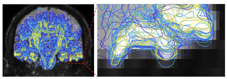
The differences between all the contours show subject movement during the scan (right).