Schilling, K. G., Combes, A. J. E., Ramadass, K., Rheault, F., Sweeney, G., Prock, L., Sriram, S., Cohen-Adad, J., Gore, J. C., Landman, B. A., Smith, S. A., & O’Grady, K. P. (2024). Influence of preprocessing, distortion correction and cardiac triggering on the quality of diffusion MR images of spinal cord. Magnetic Resonance Imaging, 108, 11–21. https://doi.org/10.1016/J.MRI.2024.01.008
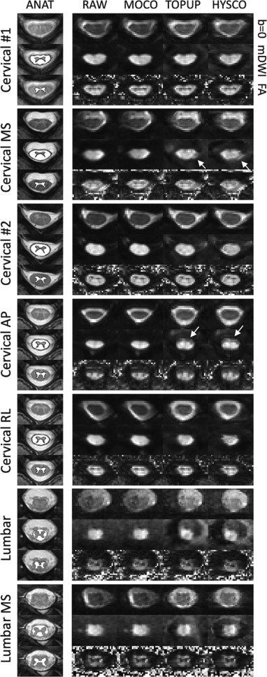
Researchers are addressing challenges in spinal cord imaging using diffusion MRI, which often suffers from issues like geometric distortion due to magnetic field irregularities and motion artifacts from biological movements. A recent study evaluated techniques to improve image quality, focusing on correcting distortions and using cardiac triggering to reduce motion effects. The findings suggest that while distortion correction methods align images better with structural scans, they do not consistently enhance the clarity or contrast within the spinal cord itself. Additionally, experiments showed that omitting cardiac triggering, which typically synchronizes image capture with heartbeats to minimize motion blur, did not degrade image quality significantly. This suggests that forgoing this technique can shorten the imaging process without sacrificing much in terms of image fidelity. The study highlights the need for further refinement in spinal cord imaging methods, particularly those adapted from brain imaging technologies.
